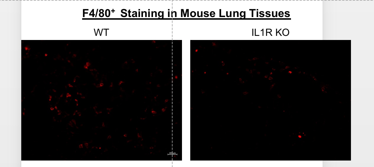
![Immunohistochemistry-Frozen: F4/80 Antibody (BM8) [PE] [NBP2-22134] - Red: F4/80, Green: CD31/PECAM-1, Blue: DAPI+ nuclei. Frozen mouse lung tissues of WT B6 and IL1R--/- (knockout) mice were blocked with 1% BSA in PBS and were double stained with PE-conjugated F4/80 and Alex647-conjugated CD31/PECAM-1 antibody (MEC 7.46) for 2 hours at the room temperature. This image was submitted via a customer review.](http://images.novusbio.com/fullsize/F4-80-Antibody-BM8-PE-Immunohistochemistry-Frozen-NBP2-22134-img0001.jpg "Immunohistochemistry-Frozen: F4/80 Antibody (BM8) [PE] [NBP2-22134] - Red: F4/80, Green: CD31/PECAM-1, Blue: DAPI+ nuclei. Frozen mouse lung tissues of WT B6 and IL1R--/- (knockout) mice were blocked with 1% BSA in PBS and were double stained with PE-conjugated F4/80 and Alex647-conjugated CD31/PECAM-1 antibody (MEC 7.46) for 2 hours at the room temperature. This image was submitted via a customer review.")
| Immunogen | This F4/80 Antibody (BM8) was developed against BALB/c macrophages obtained from 14-day-old bone marrow cell cultures. |
| Marker | Macrophage Marker |
| Isotype | IgG2a |
| Clonality | Monoclonal |
| Host | Rat |
| Gene | Adgre1 |
| Purity | Protein A or G purified |
| Innovator's Reward | Test in a species/application not listed above to receive a full credit towards a future purchase. |
| Dilutions |
|
|
| Application Notes | This antibody works in Flow Cytometry, Immunohistochemistry-Frozen, Immunohistochemistry-Paraffin, Western Blot. The typical starting working dilution is 1:50. F4/80 is sensitive to 2-mercaptoethanol. The antibody requires proteinase K treatment of paraffin sections. IHC-P and Flow reported in a verified customer review. |
|
| Reviewed Applications |
|
| Storage | Store at 4C in the dark. |
| Buffer | PBS and 1.0% BSA |
| Preservative | 0.02% Sodium Azide |
| Purity | Protein A or G purified |


![Flow Cytometry B7-1/CD80 Antibody (37711) [Unconjugated]](https://images.novusbio.com/images2/B71_MAB140_Flow_Cytometry_16025.jpg)
![Immunohistochemistry B7-1/CD80 Antibody (37711) [Unconjugated]](https://images.novusbio.com/images2/B71_MAB140_Immunohistochemistry_17363.jpg)
![Dual RNAscope ISH-IHC Compatible B7-1/CD80 Antibody (37711) [Unconjugated]](https://images.novusbio.com/images2/B71_MAB140_Dual_RNAscope_ISHIHC_23827.jpg)
![F4/80 Antibody (BM8) [PE]](/sites/all/modules/enterprise-tech/et_datasheets/images/novus_guarantee.png "F4/80 Antibody (BM8) [PE]")
| Images | Ratings | Applications | Species | Date | Details | ||||||||||
|---|---|---|---|---|---|---|---|---|---|---|---|---|---|---|---|
Enlarge |
reviewed by:
James Xiao |
IHC-Fr | Mouse | 03/02/2018 |
Summary
Comments
|
||||||||||

Enlarge |
reviewed by:
James Xiao |
Flow | Mouse | 03/02/2018 |
Summary
Comments
|
Secondary Antibodies |
Isotype Controls |
Research Areas for F4/80 Antibody (NBP2-22134)Find related products by research area.
|
|
TMEM 119 is a specific marker of microglia cells By Jennifer Sokolowski, MD, PhD.Microglia are a major immune-cell component in the brain. They ingest and degrade dead cells, debris, and foreign material and interact with other immune cells to orchestrate centra... Read full blog post. |
The concentration calculator allows you to quickly calculate the volume, mass or concentration of your vial. Simply enter your mass, volume, or concentration values for your reagent and the calculator will determine the rest.
5 | |
4 | |
3 | |
2 | |
1 |
| James Xiao 03/02/2018 |
||
| Application: | IHC-Fr | |
| Species: | Mouse |
| James Xiao 03/02/2018 |
||
| Application: | Flow | |
| Species: | Mouse |
| Gene Symbol | Adgre1 |


![Flow Cytometry: F4/80 Antibody (BM8) [PE] [NBP2-22134] - Splenocytes isolated from a IL1R-/- mouse (IL1b receptor KO) were stained with F4/80 Antibody (BM8) and CD45-e450 (1:100 in FACS buffer) for 30 minutes. Cells were then washed and analyzed by flow cytometry. Left panel shows the isotype control. This image was submitted via a customer review.](http://images.novusbio.com/fullsize/F4-80-Antibody-BM8-PE-Flow-Cytometry-NBP2-22134-img0002.jpg "Flow Cytometry: F4/80 Antibody (BM8) [PE] [NBP2-22134] - Splenocytes isolated from a IL1R-/- mouse (IL1b receptor KO) were stained with F4/80 Antibody (BM8) and CD45-e450 (1:100 in FACS buffer) for 30 minutes. Cells were then washed and analyzed by flow cytometry. Left panel shows the isotype control. This image was submitted via a customer review.")
![SDS-PAGE TNF-alpha [Unconjugated]](https://images.novusbio.com/images2/TNF-alpha_210-TA_256.jpg)
![Bioactivity TNF-alpha [Unconjugated]](https://images.novusbio.com/images2/TNFalpha_210TA_1658.jpg)
![SEC-MALS TNF-alpha [Unconjugated]](https://images.novusbio.com/images/210-ta_recombinant-human-tnf-alpha-protein-sec-mals-35202312244..jpg)
![N/A CCL2/JE/MCP-1 [HRP]](https://images.novusbio.com/images2/CCL2_DCP00_ELISA_67.jpg)
![N/A CCL2/JE/MCP-1 [HRP]](https://images.novusbio.com/images2/DATA_CCL2_DCP00_ELISA_633.jpg)
![N/A CCL2/JE/MCP-1 [HRP]](https://images.novusbio.com/images2/DATA_CCL2_DCP00_ELISA_634.jpg)
![N/A IL-6 [HRP]](https://images.novusbio.com/images2/DATA_IL6_M6000_ELISA_936.jpg)
![N/A IL-6 [HRP]](https://images.novusbio.com/images2/IL-6_M6000_ELISA_415.jpg)
![N/A IL-6 [HRP]](https://images.novusbio.com/images/m6000b_mouse-il-6-quantikine-elisa-kit-44202415433118.jpg)
![Immunocytochemistry Integrin beta 2/CD18 Antibody [Unconjugated]](https://images.novusbio.com/images2/Integrin_beta_2_AF1730_Immunocytochemistry_6474.jpg)
![Immunocytochemistry Integrin beta 2/CD18 Antibody [Unconjugated]](https://images.novusbio.com/images/antibody/af1730_human-integrin-beta-2-cd18-affinity-purified-polyclonal-ab-immunocytochemistry-1292023225343.jpg)
![Flow Cytometry Integrin beta 2/CD18 Antibody [Unconjugated]](https://images.novusbio.com/images/antibody/af1730_human-integrin-beta-2-cd18-affinity-purified-polyclonal-ab-flow-cytometry-7220258626..jpg)
![N/A IL-10 [Biotin]](https://images.novusbio.com/images2/DATA_IL10_DY417_ELISA_2014.jpg)
![Immunohistochemistry CD45 Antibody [Unconjugated]](https://images.novusbio.com/images2/CD45_AF114_Immunohistochemistry_23525.jpg)
![Immunocytochemistry CD45 Antibody [Unconjugated]](https://images.novusbio.com/images2/af114_mouse-cd45-affinity-purified-polyclonal-ab-immunocytochemistry-6122021145449.jpg)
![Immunocytochemistry/ Immunofluorescence CD45 Antibody [Unconjugated]](https://images.novusbio.com/images/af114_mouse-cd45-affinity-purified-polyclonal-ab-41202410331954.jpg)
![Bioactivity CTLA-4 [Unconjugated]](https://images.novusbio.com/images2/CTLA4_7268CT_2293.jpg)
![Bioactivity IL-4 [Unconjugated]](https://images.novusbio.com/images/protein/6507-ilcf_recombinant-human-il-4-cho-expressed-protein-cf-bioactivity-272020133214.jpg)
![Bioactivity IFN-gamma [Unconjugated]](https://images.novusbio.com/images2/IFNgamma_285IF_2848.jpg)
![SDS-PAGE IFN-gamma [Unconjugated]](https://images.novusbio.com/images2/IFN-gamma_285-IF_230.jpg)
![SEC-MALS IFN-gamma [Unconjugated]](https://images.novusbio.com/images/285-if_recombinant-human-ifn-gamma-protein-sec-mals-1520239228..jpg)


![Bioactivity GM-CSF [Unconjugated]](https://images.novusbio.com/images/protein/7954-gmcf_recombinant-human-gm-csf-cho-expressed-protein-cf-bioactivity-272020121514.jpg)

