 at 1 µg/mL at 37 ° Celsius for 4 minutes. Before incubation with the primary antibody, tissue underwent an all-in-one dewaxing and antigen retrieval preprocessing using PreTreatment Module (PT Module) and Dewax and HIER Buffer H (pH 9). Tissue was stained using the Alexa Fluor™ Plus 647 Goat anti-Rat IgG Secondary Antibody at 1:200 at 37 ° Celsius for 2 minutes. (Yellow; Lunaphore Catalog # DR647RT) and counterstained with DAPI (blue; Lunaphore Catalog # DR100). Specific staining was localized to the cytoplasm and cytoskeleton. Protocol available in COMET™ Panel Builder.")
| Reactivity | Hu, Mu, RtSpecies Glossary |
| Applications | WB, Simple Western, IHC, Flow, ICC/IF, KO, mIF |
| Clone | 280618 |
| Clonality | Monoclonal |
| Host | Rat |
| Conjugate | Unconjugated |
| Concentration | LYOPH |
| Immunogen | E. coli-derived recombinant human Vimentin Ser2-Glu466 Accession # P08670 |
| Specificity | Vimentin antibodies are ideal for immunocytochemistry colocalization studies in intermediate filaments. Detects human, mouse and rat Vimentin in Western blots. |
| Source | N/A |
| Isotype | IgG2a |
| Clonality | Monoclonal |
| Host | Rat |
| Gene | VIM |
| Purity Statement | Protein A or G purified from hybridoma culture supernatant |
| Innovator's Reward | Test in a species/application not listed above to receive a full credit towards a future purchase. |
| Dilutions |
|
|
| Reviewed Applications |
|
|
| Publications |
|
| Storage | Use a manual defrost freezer and avoid repeated freeze-thaw cycles.
|
| Buffer | Lyophilized from a 0.2 μm filtered solution in PBS with Trehalose. See Certificate of Analysis for details. *Small pack size (-SP) is supplied either lyophilized or as a 0.2 µm filtered solution in PBS. |
| Preservative | No Preservative |
| Concentration | LYOPH |
| Reconstitution Instructions | Reconstitute at 0.5 mg/mL in sterile PBS. For liquid material, refer to CoA for concentration. |
Vimentin is a 57 kDa class III intermediate filament (IF) protein that belongs to the intermediate filament family. It is the predominant IF in cells of mesenchymal origin such as vascular endothelium and blood cells (1-3). The human Vimentin cDNA encodes a 466 amino acid (aa) protein that contains head and tail regions with multiple regulatory Ser/Thr phosphorylation sites, and a central rod domain with three coiled-coil regions separated by linkers (1, 2). Human Vimentin shares 97-98% aa identity with mouse, rat, ovine, bovine, and canine Vimentin. Sixteen Vimentin coiled-coil dimers self-assemble to form intermediate (10-12 nm wide) filaments (4). These filaments then anneal longitudinally to form non-polarized fibers that support cell structure and withstand stress (4). IF fibers are highly dynamic, and half-life depends on the balance between kinase and phosphatase activity. For example, phosphorylation followed by dephosphorylation drives IF disintegration, followed by reorganization during mitosis (1, 5, 6). Interactions of head and tail domains link IFs with other structures such as actin and microtubule cytoskeletons (7). Vimentin is involved in positioning autophagosomes, lysosomes and the Golgi complex within the cell (8). It facilitates cell migration and motility by recycling internalized trailing edge integrins back to the cell surface at the leading edge (9-11). Vimentin helps maintain the lipid composition of cellular membranes, and caspase cleavage of Vimentin is a key event in apoptosis (8, 12). Phosphorylation promotes secretion of Vimentin by TNF-alpha -stimulated macrophages (13). Extracellular Vimentin has been shown to associate with several microbes, and appears to promote an antimicrobial oxidative burst (13, 14). Cell-associated Vimentin can also interact with NKp46 to recruit NK cells to tuberculosis-infected monocytes (15).

The concentration calculator allows you to quickly calculate the volume, mass or concentration of your vial. Simply enter your mass, volume, or concentration values for your reagent and the calculator will determine the rest.


 at 1 µg/mL at 37 ° Celsius for 4 minutes. Before incubation with the primary antibody, tissue underwent an all-in-one dewaxing and antigen retrieval preprocessing using PreTreatment Module (PT Module) and Dewax and HIER Buffer H (pH 9; Epredia Catalog # TA-999-DHBH). Tissue was stained using the Alexa Fluor™ 647 Goat anti-Rat IgG Secondary Antibody at 1:200 at 37 ° Celsius for 2 minutes. (Yellow; Lunaphore Catalog # DR647RT) and counterstained with DAPI (blue; Lunaphore Catalog # DR100). Specific staining was localized to the cytoplasm and cytoskeleton. Protocol available in COMET™ Panel Builder.")
 at 1 µg/mL at 37 ° Celsius for 4 minutes. Before incubation with the primary antibody, tissue underwent an all-in-one dewaxing and antigen retrieval preprocessing using PreTreatment Module (PT Module) and Dewax and HIER Buffer H (pH 9; Epredia Catalog # TA-999-DHBH). Tissue was stained using the Alexa Fluor™ 647 Goat anti-Rat IgG Secondary Antibody at 1:200 at 37 ° Celsius for 2 minutes. (Yellow; Lunaphore Catalog # DR647RT) and counterstained with DAPI (blue; Lunaphore Catalog # DR100). Specific staining was localized to the cytoplasm and cytoskeleton. Protocol available in COMET™ Panel Builder.")
 at 10ug/mL at 37 ° Celsius for 4 minutes. Before incubation with the primary antibody, tissue underwent an all-in-one dewaxing and antigen retrieval preprocessing using PreTreatment Module (PT Module) and Dewax and HIER Buffer H (pH 9; Epredia Catalog # TA-999-DHBH). Tissue was stained using the Alexa Fluor™ 647 Goat anti-Rat IgG Secondary Antibody at 1:200 at 37 ° Celsius for 2 minutes. (Yellow; Lunaphore Catalog # DR647RT) and counterstained with DAPI (blue; Lunaphore Catalog # DR100). Specific staining was localized to the membrane. Protocol available in COMET™ Panel Builder.")
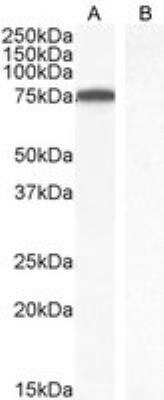
 followed by HRP-conjugated Anti-Rat IgG Secondary Antibody (Catalog # HAF005). A specific band was detected for Vimentin at approximately 55 kDa (as indicated). This experiment was conducted under reducing conditions and using Immunoblot Buffer Group 1.")
 followed by HRP-conjugated Anti-Rat IgG Secondary Antibody (Catalog # HAF005). A specific band was detected for Vimentin at approximately 55 kDa (as indicated). This experiment was conducted under reducing conditions and using Immunoblot Buffer Group 1.")
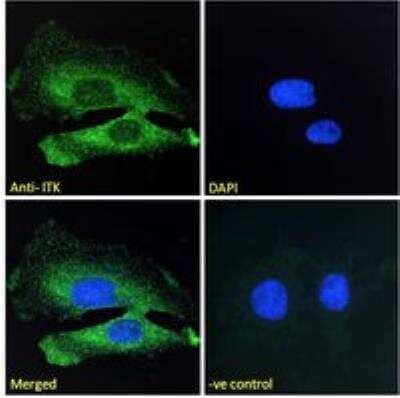
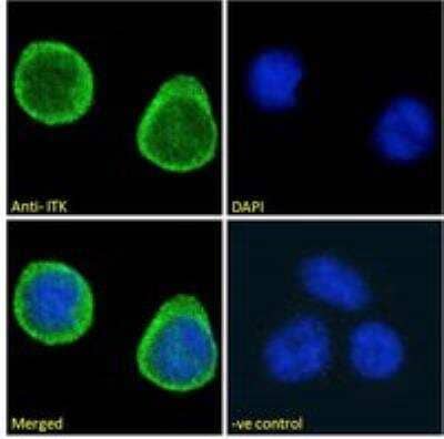
 at 10 µg/mL for 3 hours at room temperature. Cells were stained using the NorthernLights™ 557-conjugated Anti-Rat IgG Secondary Antibody (yellow; Catalog # NL013) and counterstained with DAPI (blue). View our protocol for Fluorescent ICC Staining of Cells on Coverslips.")
 at 10 µg/mL for 3 hours at room temperature. Cells were stained using the NorthernLights™ 493-conjugated Anti-Rat IgG Secondary Antibody (green; Catalog # NL015) and counterstained with DAPI (blue). View our protocol for Fluorescent ICC Staining of Cells on Coverslips.")
 at 10 µg/mL for 3 hours at room temperature. Cells were stained using the NorthernLights™ 557-conjugated Anti-Rat IgG Secondary Antibody (red; Catalog # NL013) and counterstained with DAPI (blue). Specific staining was localized to cytoskeleton. View our protocol for Fluorescent ICC Staining of Cells on Coverslips.")
 at 10 µg/mL for 3 hours at room temperature. Cells were stained using the NorthernLights™ 557-conjugated Anti-Rat IgG Secondary Antibody (red; Catalog # NL013) and counterstained with DAPI (blue). Specific staining was localized to cytoskeleton. View our protocol for Fluorescent ICC Staining of Cells on Coverslips.")
 at 0.5 µg/mL for 1 hour at room temperature followed by incubation with the Anti-Rat IgG VisUCyte™ HRP Polymer Antibody (Catalog # VC005). Tissue was stained using DAB (brown) and counterstained with hematoxylin (blue). Specific staining was localized to cytoplasm. View our protocol for IHC Staining with VisUCyte HRP Polymer Detection Reagents.")
 or isotype control antibody (Catalog # MAB006, open histogram) followed by anti-Rat IgG PE-conjugated secondary antibody (Catalog # F0105B). To facilitate intracellular staining, cells were fixed with Flow Cytometry Fixation Buffer (Catalog # FC004) and permeabilized with Flow Cytometry Permeabilization/Wash Buffer I (Catalog # FC005). View our protocol for Staining Intracellular Molecules.")
 using 10 µg/mL of Rat Anti-Human/Mouse/Rat Vimentin Monoclonal Antibody (Catalog # MAB2105) followed by 1:50 dilution of HRP-conjugated Anti-Rat IgG Secondary Antibody (Catalog # HAF005). This experiment was conducted under reducing conditions and using the 12-230 kDa separation system.")
. PVDF membrane was probed with 2 µg/mL of Rat Anti-Human/Mouse/Rat Vimentin Monoclonal Antibody (Catalog # MAB2105) followed by HRP-conjugated Anti-Goat IgG Secondary Antibody (HAF017). A specific band was detected for Vimentin at approximately 55 kDa (as indicated) in the parental K562 cell line, but is not detectable in knockout K562 cell line. GAPDH (MAB5718) is shown as a loading control. This experiment was conducted under reducing conditions and using Western Blot Buffer Group 1.")
 by vimentin+ (grey) HSCs/myofibroblasts (A) or CD31+ (grey) ECs (C). (A) The majority of vimentin+ cells were Pc-ve in the tissues examined. Representative image shown, displaying absence of Pc on vimentin+ cells. To confirm this result, ciliary protein Arl13b (green) was co-stained with vimentin (grey). Rare Arl13b ciliary structures (arrow) co-localised with vimentin+ cells. Final panel in A illustrates rare Pc+ ( alpha -acetylated tubulin, green; gamma -tubulin, red) vimentin+ (grey) HSCs/myofibroblasts, at the cirrhotic interface. (B) Number of vimentin+ Pc+ cells or vimentin+ Pcneg cells per FOV (n = 3 ALD samples, 8 FOV/sample). (C) No Pc were detected on CD31+ cells in the tissues examined (ALD n = 3, 8 FOV/sample). Representative image shown. All images obtained using confocal microscopy, 63x objective. DAPI, blue. White arrows illustrate Pc. * Non-specific liver autofluorescence. Image collected and cropped by CiteAb from the following publication (//dx.plos.org/10.1371/journal.pone.0171480), licensed under a CC-BY license. Not internally tested by R&D Systems.")
![Widespread GLI expression in human donor and cirrhotic liver.(A) Frozen (4 μm) human donor (n = 5), and cirrhotic liver sections [ALD (n = 6), NASH (n = 3), PBC (n = 1)] were screened for GLI2 (red) expression by immunofluorescence. Representative images taken at 5x or 40x (insets) objective shown. DAPI, blue. (B) qRT-PCR for GLI1 and GLI3 transcript in human donor or ALD samples. Mean±S.E.M. Significant (*) difference between means (One-sided student t-test, **p150 kDa) in donor (Don) or ALD patient samples. Densitometry analysis with GLI1 normalised to GAPDH (Image J). Mean±S.E.M; **p = 0.0093 (Two-sided student t-test). (C) Nuclear GLI2 (green) expression in EpCAM+ (red) LPCs in donor, ALD, PBC and NASH liver. (D) Nuclear GLI2 (green) expression demonstrated within CD31+ (red) ECs, CK18+ (red) hepatocytes, CD45+ (red) leukocytes and vimentin+ (red) HSCs/myofibroblasts, in ALD. 63x objective. (E) Maximum intensity projection illustrating close physical association between EpCAM+ LPCs (green) and vimentin+ HSCs/myofibroblasts (red), both of which express GLI2 (grey), in ALD tissue. Arrows indicate myofibroblasts directly contacting LPCs. Confocal microscopy, 63x objective. Quantitation (%) of EpCAM+ GLI2+ cells and vimentin+ GLI2+ cells within the same FOV (n = 3 ALD samples, 8 FOV/sample). Image collected and cropped by CiteAb from the following publication (//dx.plos.org/10.1371/journal.pone.0171480), licensed under a CC-BY license. Not internally tested by R&D Systems.](http://images.novusbio.com/fullsize/mab2105_human-mouse-rat-vimentin-mab-clone-280618-41202410331991.jpg "Widespread GLI expression in human donor and cirrhotic liver.(A) Frozen (4 μm) human donor (n = 5), and cirrhotic liver sections [ALD (n = 6), NASH (n = 3), PBC (n = 1)] were screened for GLI2 (red) expression by immunofluorescence. Representative images taken at 5x or 40x (insets) objective shown. DAPI, blue. (B) qRT-PCR for GLI1 and GLI3 transcript in human donor or ALD samples. Mean±S.E.M. Significant (*) difference between means (One-sided student t-test, **p150 kDa) in donor (Don) or ALD patient samples. Densitometry analysis with GLI1 normalised to GAPDH (Image J). Mean±S.E.M; **p = 0.0093 (Two-sided student t-test). (C) Nuclear GLI2 (green) expression in EpCAM+ (red) LPCs in donor, ALD, PBC and NASH liver. (D) Nuclear GLI2 (green) expression demonstrated within CD31+ (red) ECs, CK18+ (red) hepatocytes, CD45+ (red) leukocytes and vimentin+ (red) HSCs/myofibroblasts, in ALD. 63x objective. (E) Maximum intensity projection illustrating close physical association between EpCAM+ LPCs (green) and vimentin+ HSCs/myofibroblasts (red), both of which express GLI2 (grey), in ALD tissue. Arrows indicate myofibroblasts directly contacting LPCs. Confocal microscopy, 63x objective. Quantitation (%) of EpCAM+ GLI2+ cells and vimentin+ GLI2+ cells within the same FOV (n = 3 ALD samples, 8 FOV/sample). Image collected and cropped by CiteAb from the following publication (//dx.plos.org/10.1371/journal.pone.0171480), licensed under a CC-BY license. Not internally tested by R&D Systems.")
 at 5 µg/ml overnight at 4 °C. Before incubation with the primary antibody, tissue was subjected to heat-induced epitope retrieval using VisUCyte Antigen Retrieval Reagent-Basic (Catalog # VCTS021). Tissue was stained using the HRP-conjugated Anti-Rat IgG Secondary Antibody (Catalog # HAF005) and counterstained with hematoxylin (blue). Specific staining was localized to the cytoplasm. View our protocol for Chromogenic IHC Staining of Paraffin-embedded Tissue Sections.")
![Immunohistochemistry Desmin Antibody [Unconjugated]](https://images.novusbio.com/images2/Desmin_AF3844_Immunohistochemistry_10689.jpg)
![Immunohistochemistry Desmin Antibody [Unconjugated]](https://images.novusbio.com/images2/Desmin_AF3844_Immunohistochemistry_6808.jpg)
![Immunocytochemistry/ Immunofluorescence Desmin Antibody [Unconjugated]](https://images.novusbio.com/images/af3844_human-mouse-desmin-affinity-purified-polyclonal-ab-41202410331920.jpg)


![Immunohistochemistry CD34 Antibody [Unconjugated]](https://images.novusbio.com/images2/CD34_AF4117_Immunohistochemistry_6824.jpg)


![Immunocytochemistry E-Cadherin Antibody [Unconjugated]](https://images.novusbio.com/images2/ECadherin_AF748_Immunocytochemistry_15885.jpg)
![Immunohistochemistry E-Cadherin Antibody [Unconjugated]](https://images.novusbio.com/images2/ECadherin_AF748_Immunohistochemistry_15886.jpg)
![Immunocytochemistry E-Cadherin Antibody [Unconjugated]](https://images.novusbio.com/images2/ECadherin_AF748_Immunocytochemistry_16572.jpg)


![Simple Western CD117/c-kit Antibody [Unconjugated]](https://images.novusbio.com/images2/CD117_AF1356_Simple_Western_17480.jpg)
![Western Blot CD117/c-kit Antibody [Unconjugated]](https://images.novusbio.com/images2/CD117_AF1356_Western_Blot_18637.jpg)
![Immunohistochemistry CD117/c-kit Antibody [Unconjugated]](https://images.novusbio.com/images2/SCF_R_AF1356_Immunohistochemistry_9746.jpg)


![Vimentin Antibody (280618) [Unconjugated]](/sites/all/modules/enterprise-tech/et_datasheets/images/novus_guarantee.png "Vimentin Antibody (280618) [Unconjugated]")

 or Rat IgG Isotype Control Antibody (Catalog #MAB006,open histogram), followed by Phycoerythrin-conjugated Anti-Rat IgG SecondaryAntibody (Catalog # F0105B).")
