 and Hoechst (blue). IHC-P image submitted by a verified customer review.")
| Reactivity | Hu, Mu, Rt, Po, Bv, RbSpecies Glossary |
| Applications | WB, ELISA, ICC/IF, IHC, IP, MA, PAGE, KD |
| Clonality | Polyclonal |
| Host | Rabbit |
| Conjugate | Unconjugated |
| Format | BSA Free |
| Description | This antibody has been prepared by immunoaffinity chromatography using immobilized antigens followed by extensive cross-adsorption against other collagens, human serum proteins and non-collagen extracellular matrix proteins to remove any unwanted specificities. Some class-specific anti-collagens may be specific for three-dimensional epitopes which may result in diminished reactivity with denatured collagen or formalin-fixed, paraffin embedded tissues. Store vial at 4C prior to opening. This product is stable at 4C as an undiluted liquid. Dilute only prior to immediate use. For extended storage, mix with an equal volume of glycerol, aliquot contents and freeze at -20C or below. Avoid cycles of freezing and thawing. |
| Immunogen | Collagen I from human and bovine placenta (Uniprot: P02452) |
| Localization | Extracellular matrix |
| Specificity | Some class-specific anti-collagens may be specific for three-dimensional epitopes which may result in diminished reactivity with denatured collagen or formalin-fixed, paraffin embedded tissues. This antibody reacts with most mammalian Type I collagens and has expected cross-reactivity with Type III and negligible cross reactivity with Type II, IV, V or VI collagens. Non-specific cross-reaction of anti-collagen antibodies with other human serum proteins or non-collagen extracellular matrix proteins has not been tested. |
| Isotype | IgG |
| Clonality | Polyclonal |
| Host | Rabbit |
| Gene | COL1A1 |
| Purity | Immunogen affinity purified |
| Innovator's Reward | Test in a species/application not listed above to receive a full credit towards a future purchase. |
| Dilutions |
|
|
| Application Notes | This product has been tested by dot blot and IHC and are useful for indirect trapping ELISA for quantitation of antigen in serum using a standard curve, immunoprecipitation, native (non-denaturing, non-dissociating) PAGE, immunohistochemistry, Immunofluorescence, FLOW, and western blotting for highly sensitive qualitative analysis. Use in SDS-Page reported in scientific literature (PMID: 30914477). |
|
| Reviewed Applications |
|
|
| Publications |
|
| Storage | Store at 4C short term. For extended storage, add an equal volume of glycerol, aliquot and store at -20C or below. Avoid repeated freeze-thaw cycles. |
| Buffer | 0.02 M Potassium Phosphate, 0.15 M Sodium Chloride, pH 7.2 |
| Preservative | 0.01% Sodium Azide |
| Purity | Immunogen affinity purified |


![Immunocytochemistry Osteopontin/OPN Antibody [Unconjugated]](https://images.novusbio.com/images2/Osteopontin_AF808_Immunocytochemistry__Immunofluorescence_23325.jpg)
![Neutralization Osteopontin/OPN Antibody [Unconjugated]](https://images.novusbio.com/images2/Osteopontin_AF808_Block_Neutralize_8975.jpg)
![Immunohistochemistry Osteopontin/OPN Antibody [Unconjugated]](https://images.novusbio.com/images2/Osteopontin_AF808_Immunohistochemistry_9333.jpg)
![Immunohistochemistry PTH Antibody (918462) [Unconjugated]](https://images.novusbio.com/images2/PTH_MAB7665_Immunohistochemistry_16650.jpg)
![Western Blot PTH Antibody (918462) [Unconjugated]](https://images.novusbio.com/images2/PTH_MAB7665_Western_Blot_16656.jpg)


| Images | Ratings | Applications | Species | Date | Details | ||||||||||
|---|---|---|---|---|---|---|---|---|---|---|---|---|---|---|---|
Enlarge |
reviewed by:
Federica Gnudi |
IHC-P | Rat | 05/05/2022 |
Summary
|
||||||||||

Enlarge |
reviewed by:
Verified Customer |
ICC | Human | 04/23/2021 |
Summary
|
||||||||||

Enlarge |
reviewed by:
Verified Customer |
IHC-P | Mouse | 10/10/2019 |
Summary
|
||||||||||

Enlarge |
reviewed by:
Verified Customer |
IHC-P | Mouse | 10/02/2019 |
Summary
|
||||||||||
|
reviewed by:
Wangsheng Wang |
WB | Human | 05/30/2018 |
Summary
|
|||||||||||

Enlarge |
reviewed by:
Verified Customer |
IHC-P | Mouse | 10/25/2017 |
Summary
|
||||||||||

Enlarge |
reviewed by:
Verified Customer |
WB | Human | 09/27/2017 |
Summary
|
||||||||||
|
reviewed by:
Verified Customer |
ICC | Feline | 06/23/2017 |
Summary
Comments
|
|||||||||||

Enlarge |
reviewed by:
Jakob Townsend |
IHC-P | Rat | 02/20/2017 |
Summary
|
||||||||||

Enlarge |
reviewed by:
Verified Customer |
IHC-P | Rat | 07/20/2016 |
Summary
|
||||||||||

Enlarge |
reviewed by:
Verified Customer |
WB | Other | 08/04/2015 |
Summary
|
||||||||||

Enlarge |
reviewed by:
Verified Customer |
IF | Human | 10/02/2014 |
Summary
|
||||||||||

Enlarge |
reviewed by:
Elizabeth Rendina-Ruedy |
WB | Mouse | 08/05/2013 |
Summary
|
||||||||||

Enlarge |
reviewed by:
Verified Customer |
IHC-Fr | Human | 06/18/2013 |
Summary
|
||||||||||

Enlarge |
reviewed by:
Nathan Weidenhamer |
ICC | Human | 02/04/2013 |
Summary
|
||||||||||
.jpg)
Enlarge |
reviewed by:
Verified Customer |
WB | Human | 09/04/2012 |
Summary
|
Secondary Antibodies |
Isotype Controls |
Research Areas for Collagen I Antibody (NB600-408)Find related products by research area.
|
The concentration calculator allows you to quickly calculate the volume, mass or concentration of your vial. Simply enter your mass, volume, or concentration values for your reagent and the calculator will determine the rest.
5 | |
4 | |
3 | |
2 | |
1 |
| Federica Gnudi 05/05/2022 |
||
| Application: | IHC-P | |
| Species: | Rat |
| Verified Customer 04/23/2021 |
||
| Application: | ICC | |
| Species: | Human |
| Verified Customer 10/10/2019 |
||
| Application: | IHC-P | |
| Species: | Mouse |


 and COLLAGEN 1 (red) immunostaining with DAPI (blue) in non-treated (control), TGFbeta-treated cells with or without gamma-secretase inhibitor. Scale bars: 50 um Image collected and cropped by CiteAb from the following publication (//www.nature.com/articles/s41467-019-09992-3) licensed under a CC-BY license.")
: Collagen Type I splice variants and isoforms.")
 licensed under a CC-BY license.")
, CD34, and DAPI. The position of the septa is underlined, scale bar: 100 um. Image collected and cropped by CiteAb from the following publication (//www.nature.com/articles/s41467-019-09992-3) licensed under a CC-BY license.")


 Collagen I (red) and fibronectin (blue) staining of patient #1 and #3 NF and CAF CDM. Scale bar, 20 μm. Image collected and cropped by CiteAb from the following publication (//www.nature.com/doifinder/10.1038/ncomms12237) licensed under a CC-BY license.")
 frozen tumor sections. Image provided by Dr. Wa'el Al Rawashdeh of RWTH Aachen University.")
 alpha-smooth muscle actin NBP2-34760APC (yellow), CD45 (red) and Collagen I NB600-408 (top) or Collagen III/COL3A1 NB600-594 (bottom) (blue) in the liver of uninfected or 9 wpi WT mice. Scale bars represent 50 um. Image collected and cropped by CiteAb from the following publication (//pubmed.ncbi.nlm.nih.gov/32973293/) licensed under a CC-BY license.")

 in control (GFP-transduced) (left lane) and PPARg-transduced cell lysates (right lane). Protein staining shown below each blot depicts equal protein loading. An equal amount of the whole cell protein (100 ug) was separated by SDS-PAGE and electroblotted to nitro-cellulose membranes. Proteins were detected by incubating the membrane with anti-Collagen I antibody at a concentration of 0.2-2 ug/10 mL in TBS (100 mM Tris-HCl, 0.15 M NaCl, pH 7.4) with 5% non-fat milk. Detection occurred by incubation with a HRP conjugated secondary antibody at 1 ug/10 mL. Proteins were detected by a chemiluminescent method using the PIERCE ECL kit (Amersham Biosciences).")


 fibrosis, B: Non-fibrotic CV, C: Perisinusodial fibrosis, D: Non-fibrotic area, E: Protat tract fibrosis, F: Septal fibrosis (arrow). Antigen retrieval: not required. Primary antibody: Anti-collagen type I at 1:1250 for 4C for 24hr. Secondary antibody: Peroxidase biotin-streptavidin rabbit secondary antibody at 1:10,000 for 45 min at RT. Localization: Anti-collagen type I is intra and extracellular. Staining: 3.3'-diaminobenzidine tetrahydrochloride was used as the chromogen. Nuclei were counterstained purple with hematoxylin.")


 with hematoxylin purple nuclear counterstain. With corresponding negative conrol (B).")

, licensed under a CC-BY license.")


 fibrosis, B: Non-fibrotic CV, C: Perisinusodial fibrosis, D: Non-fibrotic area, E: Protat tract fibrosis, F: Septal fibrosis (arrow). Fixation: formalin fixed paraffin embedded. Antigen retrieval: not required. Primary antibody: Anti-collagen type I at 1:1250 for 4C for 24hr. Secondary antibody: Peroxidase biotin-streptavidin rabbit secondary antibody at 1:10,000 for 45 min at RT. Localization: Anti-collagen type I is intra and extracellular. Staining: 3.3'-diaminobenzidine tetrahydrochloride was used as the chromogen. Nuclei were counterstained purple with hematoxylin.")
![Immunohistochemistry-Paraffin: Rabbit Polyclonal Collagen I Antibody [NB600-408] - Collagen I was stained in human FFPE tissue. Primary antibody dilution: 1/1000 in blocking buffer. HIER antigen retrieval at pH 9 for 20min. AF750 conjugated version of the antibody was used (Catalog # NB600-408AF750). Image from a verified customer review.](http://images.novusbio.com/fullsize/antibody/nb600-408_rabbit-polyclonal-collagen-i-antibody-immunohistochemistry-paraffin-1122023154839..jpg "Immunohistochemistry-Paraffin: Rabbit Polyclonal Collagen I Antibody [NB600-408] - Collagen I was stained in human FFPE tissue. Primary antibody dilution: 1/1000 in blocking buffer. HIER antigen retrieval at pH 9 for 20min. AF750 conjugated version of the antibody was used (Catalog # NB600-408AF750). Image from a verified customer review.")
 Pancreatic tumor tissues from vehicle, gemcitabine, or combination of gemcitabine plus fasudil treated KPC mice were harvested and stained for various stromal markers. Representative images are shown of H&E staining, alpha -SMA, Desmin, CD31, Collagen I, and Movat's pentachrome staining.")
 and collagen 2 (D and E) among the anterior horn, the body and the posterior horn: (A and D) young model; (B and E) adult model. Comparison of collagen 1 (C) and collagen 2 (F) content between the young and the adult specimens. Values with different superscripts differ for P < 0.01 (A,B). (G) Representative Western blot image for collagen 1, collagen 2 and GAPDH. N/group = 8.")
![EPS15 is targeted via a SPOP/SPOPL binding consensus motif.(A) Cartoon of human EPS15 domain-organization and the amino-acid sequence. Indicated by color code are the SPOP/SPOPL binding site (red) and the lysine residue (yellow), which is ubiquitinated in a CRL3SPOPL–dependent manner in vivo. In addition, the amino-terminal Ca2+-binding EF-hand motifs (EH), the coiled-coil domain involved in dimerization and the two carboxy-terminal ubiquitin-interacting motifs (UIMs) involved in ubiquitin-binding are indicated. (B) EPS15 ubiquitin-profiling. Peptides containing EPS15 modification sites were quantified with LC-MS/MS after enrichment of the K-epsilon -GG motif from whole cell digests of HeLa cells treated with siSPOPL or siControl. Normalized precursor mass intensity profiles for EPS15 sites corresponding to K793, K801 and K693 are shown (raw data in Figure 4—figure supplement 1B). Quantification of the beta -Actin K113 and the polyubiquitin K11 linkage peptide control for comparable enrichment. Data are mean ± SD, N = 3. **p≤0.01. (C) Purified SPOPL was incubated as indicated with GST-tagged wild-type EPS15 or GST-EPS15 mutants, where the predicted SPOPL binding motifs have been mutated individually (GST-EPS15S605-S607A and EPS15S744-S746A, respectively), pulled down with glutathione sepharose (IP [GST]) and bound proteins were analyzed by Coomassie blue staining (upper panel) and immunoblotting (lower panels). Note that SPOPL readily binds to GST-EPS15 and GST-EPS15S605-S607A, but this interaction is strongly reduced with the GST-EPS15S744-S746A mutant. (D) HeLa cells stably expressing GFP-tagged wild-type EPS15, the EPS15S744-S746A or the EPS15K793R mutants from a doxycycline-inducible promoter were transfected as indicated (+) with control siRNA or siRNA depleting SPOPL. The levels of EPS15-GFP, EGFR and for control tubulin (TUB) were analyzed by immunoblotting with specific antibodies. Experiments were quantified in Fiji and the EPS15 levels plotted as fold-increase compared to controls. Data are mean ± SEM, N = 4. *p≤0.05. Note that SPOPL depletion does not further increase the levels of both EPS15 mutants. (E) Total cell extracts were prepared from HeLa cells expressing either GFP-tagged wild-type, the EPS15S744-S746A mutant or the EPS15K793R mutant in the presence (+) or absence (-) of HA-tagged SPOPL overexpression. The levels of EPS15-GFP, SPOPL-HA and control GAPDH were analyzed by immunoblotting. Note that overexpression of SPOPL-HA is able to induce degradation of wild-type but not the EPS15S744-S746A-GFP or the EPS15K793R-GFP mutant.DOI://dx.doi.org/10.7554/eLife.13841.009EPS15 is targeted via a SPOP/SPOPL binding consensus motif.(A) Alignments of the carboxy-terminal domains of EPS15 proteins from various species. Conserved SPOPL-binding motifs and putative ubiquitination sites are highlighted by yellow boxes. (B) Peptides containing EPS15 modification sites were quantified with LC-MS/MS after enrichment of the K-epsilon -GG motif from whole cell HeLa digests treated with siSPOPL and siControl. Raw intensities for each of the triplicate LC-MS/MS runs are shown with each of the siControl conditions scaled to 100% intensity. Normalized precursor mass intensity profiles for EPS15 sites corresponding to K793, K801 and K693 are shown, with only K793 showing significant downregulation in the depletion condition. Quantification of a peptide corresponding to beta -Actin K113 and the poly-ubiquitin K11 linkage peptide is also shown to demonstrate that enrichment variations did not influence the quantification of the EPS15 sites. Additionally, the total ion chromatographic intensities for each run are plotted to provide insight into the consistency of each of the separate experiments performed on different days. Data are mean ± SD, N = 3. (C) HeLa cell lines stably expressing wild-type EPS15-GFP, the EPS15S744-746A-GFP mutant or the EPS15K793R-GFP mutant from the inducible doxycycline-promoter were treated with doxycycline for 3 days, and analyzed by live cell imaging. Displayed are maximal projections of Z-stack acquisitions, fully covering cell height. Scale bar = 10 μm.DOI://dx.doi.org/10.7554/eLife.13841.010 Image collected and cropped by CiteAb from the following open publication (//elifesciences.org/articles/13841), licensed under a CC-BY license. Not internally tested by Novus Biologicals.](http://images.novusbio.com/fullsize/nb600-408_rabbit-collagen-i-pab-23220248194310.jpg "EPS15 is targeted via a SPOP/SPOPL binding consensus motif.(A) Cartoon of human EPS15 domain-organization and the amino-acid sequence. Indicated by color code are the SPOP/SPOPL binding site (red) and the lysine residue (yellow), which is ubiquitinated in a CRL3SPOPL–dependent manner in vivo. In addition, the amino-terminal Ca2+-binding EF-hand motifs (EH), the coiled-coil domain involved in dimerization and the two carboxy-terminal ubiquitin-interacting motifs (UIMs) involved in ubiquitin-binding are indicated. (B) EPS15 ubiquitin-profiling. Peptides containing EPS15 modification sites were quantified with LC-MS/MS after enrichment of the K-epsilon -GG motif from whole cell digests of HeLa cells treated with siSPOPL or siControl. Normalized precursor mass intensity profiles for EPS15 sites corresponding to K793, K801 and K693 are shown (raw data in Figure 4—figure supplement 1B). Quantification of the beta -Actin K113 and the polyubiquitin K11 linkage peptide control for comparable enrichment. Data are mean ± SD, N = 3. **p≤0.01. (C) Purified SPOPL was incubated as indicated with GST-tagged wild-type EPS15 or GST-EPS15 mutants, where the predicted SPOPL binding motifs have been mutated individually (GST-EPS15S605-S607A and EPS15S744-S746A, respectively), pulled down with glutathione sepharose (IP [GST]) and bound proteins were analyzed by Coomassie blue staining (upper panel) and immunoblotting (lower panels). Note that SPOPL readily binds to GST-EPS15 and GST-EPS15S605-S607A, but this interaction is strongly reduced with the GST-EPS15S744-S746A mutant. (D) HeLa cells stably expressing GFP-tagged wild-type EPS15, the EPS15S744-S746A or the EPS15K793R mutants from a doxycycline-inducible promoter were transfected as indicated (+) with control siRNA or siRNA depleting SPOPL. The levels of EPS15-GFP, EGFR and for control tubulin (TUB) were analyzed by immunoblotting with specific antibodies. Experiments were quantified in Fiji and the EPS15 levels plotted as fold-increase compared to controls. Data are mean ± SEM, N = 4. *p≤0.05. Note that SPOPL depletion does not further increase the levels of both EPS15 mutants. (E) Total cell extracts were prepared from HeLa cells expressing either GFP-tagged wild-type, the EPS15S744-S746A mutant or the EPS15K793R mutant in the presence (+) or absence (-) of HA-tagged SPOPL overexpression. The levels of EPS15-GFP, SPOPL-HA and control GAPDH were analyzed by immunoblotting. Note that overexpression of SPOPL-HA is able to induce degradation of wild-type but not the EPS15S744-S746A-GFP or the EPS15K793R-GFP mutant.DOI://dx.doi.org/10.7554/eLife.13841.009EPS15 is targeted via a SPOP/SPOPL binding consensus motif.(A) Alignments of the carboxy-terminal domains of EPS15 proteins from various species. Conserved SPOPL-binding motifs and putative ubiquitination sites are highlighted by yellow boxes. (B) Peptides containing EPS15 modification sites were quantified with LC-MS/MS after enrichment of the K-epsilon -GG motif from whole cell HeLa digests treated with siSPOPL and siControl. Raw intensities for each of the triplicate LC-MS/MS runs are shown with each of the siControl conditions scaled to 100% intensity. Normalized precursor mass intensity profiles for EPS15 sites corresponding to K793, K801 and K693 are shown, with only K793 showing significant downregulation in the depletion condition. Quantification of a peptide corresponding to beta -Actin K113 and the poly-ubiquitin K11 linkage peptide is also shown to demonstrate that enrichment variations did not influence the quantification of the EPS15 sites. Additionally, the total ion chromatographic intensities for each run are plotted to provide insight into the consistency of each of the separate experiments performed on different days. Data are mean ± SD, N = 3. (C) HeLa cell lines stably expressing wild-type EPS15-GFP, the EPS15S744-746A-GFP mutant or the EPS15K793R-GFP mutant from the inducible doxycycline-promoter were treated with doxycycline for 3 days, and analyzed by live cell imaging. Displayed are maximal projections of Z-stack acquisitions, fully covering cell height. Scale bar = 10 μm.DOI://dx.doi.org/10.7554/eLife.13841.010 Image collected and cropped by CiteAb from the following open publication (//elifesciences.org/articles/13841), licensed under a CC-BY license. Not internally tested by Novus Biologicals.")
 Lymphatic vessel expansion (Lyve-1+) & HEV elongation (MECA79+) were similar between all groups. (A-ii,iii) Dense staining of collagen I & PDPN was seen in DLNs harvested from control & GelMA group compared to those from GelMA/anti-IL-6 group. (representative images from 4 different mice per group). Image collected & cropped by CiteAb from the following publication (//pubmed.ncbi.nlm.nih.gov/31024011), licensed under a CC-BY license. Not internally tested by Novus Biologicals.")
 or with a high fat & high sucrose diet (HFHSD) for 16 weeks. Mice were studied in the fed state. a Representative western blot analysis showing the contribution of Phf2 overexpression to the regulation of the PI3K/Akt signaling & pro-fibrogenic pathways in liver extracts (n = 15 per group). b (Left) Oral glucose tolerance test & insulin levels during the OGTT test. (right) Insulin tolerance test (n = 15 per group). c Liver sections stained with hematoxylin & eosin (H&E), trichrome masson & sirius red are shown. Scale bars = 100 μm (n = 10 per group). d Percentage of fibrotic area & percentage of apoptotic hepatocytes (n = 15 per group). e Relative oxidized GSSG content & measurement of Gpx activity (n = 10 per group). f Levels of carbonylated proteins (n = 10 mice per group). All error bars represent mean ± SEM. Statistical analyses were made using unpaired t-test (b) or Anova, followed by Bonferonni’s test (d, e). *P < 0.01 HFHSD/GFP compared to HFHSD/Phf2, $P < 0.01 HFHSD/GFP compared to NCD/GFP, £P < 0.01 HFHSD/Phf2 compared to HFHSD/GFP Image collected & cropped by CiteAb from the following publication (//www.nature.com/articles/s41467-018-04361-y), licensed under a CC-BY license. Not internally tested by Novus Biologicals.")
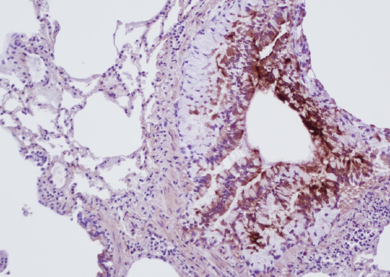
![Immunohistochemistry: Collagen I Antibody [NB600-408] - alpha -SMA, LOX, & COL1A1 immunostaining in human thyroid cancers. Representative thyroid tumors serial sections from two different patients stained by IHC for alpha -SMA, COL1A1, & LOX protein expression & localization. In the top panel, the tumor edge/invasive front is specifically shown (scale bar 500 µm), while lower panel has higher magnification (scale bar 200 µm). * clusters of tumor invading cells detaching from the principal tumor mass (T). Image collected & cropped by CiteAb from the following publication (//pubmed.ncbi.nlm.nih.gov/31906302), licensed under a CC-BY license. Not internally tested by Novus Biologicals.](http://images.novusbio.com/fullsize/nb600-408_rabbit-collagen-i-pab-310202415363722.jpg "Immunohistochemistry: Collagen I Antibody [NB600-408] - alpha -SMA, LOX, & COL1A1 immunostaining in human thyroid cancers. Representative thyroid tumors serial sections from two different patients stained by IHC for alpha -SMA, COL1A1, & LOX protein expression & localization. In the top panel, the tumor edge/invasive front is specifically shown (scale bar 500 µm), while lower panel has higher magnification (scale bar 200 µm). * clusters of tumor invading cells detaching from the principal tumor mass (T). Image collected & cropped by CiteAb from the following publication (//pubmed.ncbi.nlm.nih.gov/31906302), licensed under a CC-BY license. Not internally tested by Novus Biologicals.")
![Immunocytochemistry/ Immunofluorescence: Collagen I Antibody [NB600-408] - Increased co-localization of collagen type I with ER stress marker, KDEL in post-mortem human glaucomatous TM tissues. Post-mortem human TM tissues from age-matched normal (n = 3) & glaucoma (n = 3) were stained with Col I (green) & KDEL (red) & co-localization was examined in anterior segment tissues. Representative images are shown at lower (A) scale bar is 100 microns) & higher (B) scale bar is 25 microns) magnification. Both Col I & KDEL was increased in human glaucomatous TM tissues. In addition, increased co-localization of Col I with KDEL was observed in human glaucomatous TM tissues. Image collected & cropped by CiteAb from the following publication (//www.nature.com/articles/s41598-017-14938-0), licensed under a CC-BY license. Not internally tested by Novus Biologicals.](http://images.novusbio.com/fullsize/nb600-408_rabbit-collagen-i-pab-31020241529332.jpg "Immunocytochemistry/ Immunofluorescence: Collagen I Antibody [NB600-408] - Increased co-localization of collagen type I with ER stress marker, KDEL in post-mortem human glaucomatous TM tissues. Post-mortem human TM tissues from age-matched normal (n = 3) & glaucoma (n = 3) were stained with Col I (green) & KDEL (red) & co-localization was examined in anterior segment tissues. Representative images are shown at lower (A) scale bar is 100 microns) & higher (B) scale bar is 25 microns) magnification. Both Col I & KDEL was increased in human glaucomatous TM tissues. In addition, increased co-localization of Col I with KDEL was observed in human glaucomatous TM tissues. Image collected & cropped by CiteAb from the following publication (//www.nature.com/articles/s41598-017-14938-0), licensed under a CC-BY license. Not internally tested by Novus Biologicals.")
![Western Blot: Collagen I Antibody [NB600-408] - Expression of osteoblastic differentiation markers in Human Gingival Mesenchymal Stem Cells. Adherent HGMSCs were cultured for 21 days in control medium or in differentiation medium (Diff) supplemented or not with 1 μM resveratrol (RV). At the end, cell homogenates were processed for western blotting analysis of the expression of the osteoblastic transcription factor RUNX-2 & of the osteogenic differentiation markers COL1A1, OPN & OCN. Densitometry of the specific bands (average ± S.D.) of three independent experiments is shown in the histograms Image collected & cropped by CiteAb from the following publication (//pubmed.ncbi.nlm.nih.gov/31426798), licensed under a CC-BY license. Not internally tested by Novus Biologicals.](http://images.novusbio.com/fullsize/nb600-408_rabbit-collagen-i-pab-310202415382429.jpg "Western Blot: Collagen I Antibody [NB600-408] - Expression of osteoblastic differentiation markers in Human Gingival Mesenchymal Stem Cells. Adherent HGMSCs were cultured for 21 days in control medium or in differentiation medium (Diff) supplemented or not with 1 μM resveratrol (RV). At the end, cell homogenates were processed for western blotting analysis of the expression of the osteoblastic transcription factor RUNX-2 & of the osteogenic differentiation markers COL1A1, OPN & OCN. Densitometry of the specific bands (average ± S.D.) of three independent experiments is shown in the histograms Image collected & cropped by CiteAb from the following publication (//pubmed.ncbi.nlm.nih.gov/31426798), licensed under a CC-BY license. Not internally tested by Novus Biologicals.")
![Immunocytochemistry/ Immunofluorescence: Collagen I Antibody [NB600-408] - Dex induced ECM synthesis is associated with ER stress in TM cells. Primary human TM cells (n = 3 strains) were treated with Veh or Dex (100 nM) for 3 days & stained for KDEL & fibronectin (A) or Col I (B). A prominent co-localization of fibronectin or Col I with KDEL was observed in Dex-treated TM cells indicating induction of ER stress & excessive intracellular ECM overload in primary TM cells. (C) GTM3 cells were treated with Veh or Dex for 3 days & total lysates were subjected to immunoprecipitation using KDEL antibody & immunoblotted for ECM proteins, GRP78 & GRP94. IgG antibody was used as a negative control. Total cellular lysates were used as input. Image collected & cropped by CiteAb from the following publication (//www.nature.com/articles/s41598-017-14938-0), licensed under a CC-BY license. Not internally tested by Novus Biologicals.](http://images.novusbio.com/fullsize/nb600-408_rabbit-collagen-i-pab-310202415304285.jpg "Immunocytochemistry/ Immunofluorescence: Collagen I Antibody [NB600-408] - Dex induced ECM synthesis is associated with ER stress in TM cells. Primary human TM cells (n = 3 strains) were treated with Veh or Dex (100 nM) for 3 days & stained for KDEL & fibronectin (A) or Col I (B). A prominent co-localization of fibronectin or Col I with KDEL was observed in Dex-treated TM cells indicating induction of ER stress & excessive intracellular ECM overload in primary TM cells. (C) GTM3 cells were treated with Veh or Dex for 3 days & total lysates were subjected to immunoprecipitation using KDEL antibody & immunoblotted for ECM proteins, GRP78 & GRP94. IgG antibody was used as a negative control. Total cellular lysates were used as input. Image collected & cropped by CiteAb from the following publication (//www.nature.com/articles/s41598-017-14938-0), licensed under a CC-BY license. Not internally tested by Novus Biologicals.")
![Immunocytochemistry/ Immunofluorescence: Collagen I Antibody [NB600-408] - Dex increases Col I in the TM: Dex increases Col I & alpha -smooth muscle actin in the TM of Dex-treated mice (n = 3) compared with Veh-treated mice (n = 3). White box indicates TM. CB = ciliary body, I = Iris, C = cornea. Scale bar = 50 μm. Image collected & cropped by CiteAb from the following publication (//www.nature.com/articles/s41598-017-14938-0), licensed under a CC-BY license. Not internally tested by Novus Biologicals.](http://images.novusbio.com/fullsize/nb600-408_rabbit-collagen-i-pab-310202415175249.jpg "Immunocytochemistry/ Immunofluorescence: Collagen I Antibody [NB600-408] - Dex increases Col I in the TM: Dex increases Col I & alpha -smooth muscle actin in the TM of Dex-treated mice (n = 3) compared with Veh-treated mice (n = 3). White box indicates TM. CB = ciliary body, I = Iris, C = cornea. Scale bar = 50 μm. Image collected & cropped by CiteAb from the following publication (//www.nature.com/articles/s41598-017-14938-0), licensed under a CC-BY license. Not internally tested by Novus Biologicals.")
![Immunohistochemistry: Collagen I Antibody [NB600-408] - alpha -SMA, LOX, & COL1A1 immunostaining in human thyroid cancers. Representative thyroid tumors serial sections from two different patients stained by IHC for alpha -SMA, COL1A1, & LOX protein expression & localization. In the top panel, the tumor edge/invasive front is specifically shown (scale bar 500 µm), while lower panel has higher magnification (scale bar 200 µm). * clusters of tumor invading cells detaching from the principal tumor mass (T). Image collected & cropped by CiteAb from the following publication (//pubmed.ncbi.nlm.nih.gov/31906302), licensed under a CC-BY license. Not internally tested by Novus Biologicals.](http://images.novusbio.com/fullsize/nb600-408_rabbit-collagen-i-pab-310202415175240.jpg "Immunohistochemistry: Collagen I Antibody [NB600-408] - alpha -SMA, LOX, & COL1A1 immunostaining in human thyroid cancers. Representative thyroid tumors serial sections from two different patients stained by IHC for alpha -SMA, COL1A1, & LOX protein expression & localization. In the top panel, the tumor edge/invasive front is specifically shown (scale bar 500 µm), while lower panel has higher magnification (scale bar 200 µm). * clusters of tumor invading cells detaching from the principal tumor mass (T). Image collected & cropped by CiteAb from the following publication (//pubmed.ncbi.nlm.nih.gov/31906302), licensed under a CC-BY license. Not internally tested by Novus Biologicals.")
![Western Blot: Collagen I Antibody [NB600-408] - Spautin-1 prevents the expression of osteoblastic differentiation markers in Human Gingival Mesenchymal Stem cells. HGMSCs were cultured for 21 days in control medium or in differentiation medium supplemented with 1 μM resveratrol (Diff + RV) in the absence or in the presence of spautin-1 (Sp1). At the end, cell homogenates were processed for western blotting analysis of the expression of the osteoblastic transcription factor RUNX-2 & of the osteogenic differentiation markers COL1A1, OPN & OCN. Densitometry of the specific bands (average ± S.D.) of three independent experiments is shown in the histograms Image collected & cropped by CiteAb from the following publication (//pubmed.ncbi.nlm.nih.gov/31426798), licensed under a CC-BY license. Not internally tested by Novus Biologicals.](http://images.novusbio.com/fullsize/nb600-408_rabbit-collagen-i-pab-3102024153975.jpg "Western Blot: Collagen I Antibody [NB600-408] - Spautin-1 prevents the expression of osteoblastic differentiation markers in Human Gingival Mesenchymal Stem cells. HGMSCs were cultured for 21 days in control medium or in differentiation medium supplemented with 1 μM resveratrol (Diff + RV) in the absence or in the presence of spautin-1 (Sp1). At the end, cell homogenates were processed for western blotting analysis of the expression of the osteoblastic transcription factor RUNX-2 & of the osteogenic differentiation markers COL1A1, OPN & OCN. Densitometry of the specific bands (average ± S.D.) of three independent experiments is shown in the histograms Image collected & cropped by CiteAb from the following publication (//pubmed.ncbi.nlm.nih.gov/31426798), licensed under a CC-BY license. Not internally tested by Novus Biologicals.")
![Immunohistochemistry: Collagen I Antibody [NB600-408] - Effects of fasudil treatment on tumor stroma in KPC mice.A) Pancreatic tumor tissues from vehicle, gemcitabine, or combination of gemcitabine plus fasudil treated KPC mice were harvested & stained for various stromal markers. Representative images are shown of H&E staining, alpha -SMA, Desmin, CD31, Collagen I, & Movat's pentachrome staining. Image collected & cropped by CiteAb from the following publication (//pubmed.ncbi.nlm.nih.gov/28841710), licensed under a CC-BY license. Not internally tested by Novus Biologicals.](http://images.novusbio.com/fullsize/nb600-408_rabbit-collagen-i-pab-310202415392528.jpg "Immunohistochemistry: Collagen I Antibody [NB600-408] - Effects of fasudil treatment on tumor stroma in KPC mice.A) Pancreatic tumor tissues from vehicle, gemcitabine, or combination of gemcitabine plus fasudil treated KPC mice were harvested & stained for various stromal markers. Representative images are shown of H&E staining, alpha -SMA, Desmin, CD31, Collagen I, & Movat's pentachrome staining. Image collected & cropped by CiteAb from the following publication (//pubmed.ncbi.nlm.nih.gov/28841710), licensed under a CC-BY license. Not internally tested by Novus Biologicals.")
![Immunocytochemistry/ Immunofluorescence: Collagen I Antibody [NB600-408] - Immunofluorescence staining of osteoblastic differentiation markers in Human Gingival Mesenchymal Stem Cells. HGMSCs were plated on sterile coverslips, let adhere & cultured for 21 days in control medium or in differentiation medium (Diff) supplemented or not with 1 μM resveratrol (RV). At the end, the coverslips were fixed & processed for immunofluorescence staining of the osteogenic differentiation markers. Fluorescence staining was quantified with the ImageJ software Image collected & cropped by CiteAb from the following publication (//pubmed.ncbi.nlm.nih.gov/31426798), licensed under a CC-BY license. Not internally tested by Novus Biologicals.](http://images.novusbio.com/fullsize/nb600-408_rabbit-collagen-i-pab-31020241535614.jpg "Immunocytochemistry/ Immunofluorescence: Collagen I Antibody [NB600-408] - Immunofluorescence staining of osteoblastic differentiation markers in Human Gingival Mesenchymal Stem Cells. HGMSCs were plated on sterile coverslips, let adhere & cultured for 21 days in control medium or in differentiation medium (Diff) supplemented or not with 1 μM resveratrol (RV). At the end, the coverslips were fixed & processed for immunofluorescence staining of the osteogenic differentiation markers. Fluorescence staining was quantified with the ImageJ software Image collected & cropped by CiteAb from the following publication (//pubmed.ncbi.nlm.nih.gov/31426798), licensed under a CC-BY license. Not internally tested by Novus Biologicals.")
![Western Blot: Collagen I Antibody [NB600-408] - Collagen secretion defects in fibroblasts from patient 1a. (a) Control or patient fibroblasts incubated for 30 min to promote procollagen secretion were immunolabeled to detect COL1A1 (red) & in green, either Sec31A (to mark COPII‐coated ER exit sites) or GM130 (to mark the Golgi apparatus). (b) Extracellular collagen matrix was labeled to detect COL1A1. (c) Quantification of the mean Corrected Total Cell Fluorescence (CTCF) for COL1A1. Points show mean intensity from three independent experiments. Bars show standard deviation. (d) Immunoblot for COL1A1 in media (M) or lysates (L) of control or patient fibroblasts incubated in the presence of absence of ascorbate. (e) Unbiased proteomics was used to measure extracellular matrix collagens secreted from control or patient fibroblasts. Results from three independent experiments are shown. A ratio <1 indicates reduced collagen secretion from patient cells Image collected & cropped by CiteAb from the following publication (//pubmed.ncbi.nlm.nih.gov/31568717), licensed under a CC-BY license. Not internally tested by Novus Biologicals.](http://images.novusbio.com/fullsize/nb600-408_rabbit-collagen-i-pab-310202416163746.jpg "Western Blot: Collagen I Antibody [NB600-408] - Collagen secretion defects in fibroblasts from patient 1a. (a) Control or patient fibroblasts incubated for 30 min to promote procollagen secretion were immunolabeled to detect COL1A1 (red) & in green, either Sec31A (to mark COPII‐coated ER exit sites) or GM130 (to mark the Golgi apparatus). (b) Extracellular collagen matrix was labeled to detect COL1A1. (c) Quantification of the mean Corrected Total Cell Fluorescence (CTCF) for COL1A1. Points show mean intensity from three independent experiments. Bars show standard deviation. (d) Immunoblot for COL1A1 in media (M) or lysates (L) of control or patient fibroblasts incubated in the presence of absence of ascorbate. (e) Unbiased proteomics was used to measure extracellular matrix collagens secreted from control or patient fibroblasts. Results from three independent experiments are shown. A ratio <1 indicates reduced collagen secretion from patient cells Image collected & cropped by CiteAb from the following publication (//pubmed.ncbi.nlm.nih.gov/31568717), licensed under a CC-BY license. Not internally tested by Novus Biologicals.")
![Western Blot: Collagen I Antibody [NB600-408] - The effects of PA on TGF-beta 1-reduced PPAR gamma expression in HSC-T6 cells. (A,B) The effect of PA-induced PPAR gamma expression in rat hepatic stellate cells (HSCs). Cells were treated with PA (2 μM) for 0, 6, 12, & 24 h or PA (0.5, 1, & 2 μM) for 24 h, & PPAR gamma expression was analyzed by Western blotting. The effect of PA on TGF-beta 1-reduced PPAR gamma expression in rat HSCs; (C) cells were pretreated with various concentrations of PA (0.5, 1, & 2 μM) for 1 h, & then stimulated with TGF-beta 1 (5 ng/mL) for 24 h. Total protein extracted from cells was subjected to Western blotting to determine PPAR gamma expression; (D,E) cells were treated with TGF-beta 1 (5 ng/mL) for 24 h in the presence of PA and/or GW9662 (10 μM). Total protein extracted from cells was subjected to Western blotting to determine the expression of PPAR gamma & alpha -SMA & collagen I alpha 1. Protein bands were imaged using densitometry & analyzed using ImageJ software. The relative expression levels of target proteins were normalized using beta -actin as an internal control. The results are presented as the means ± SD of three independent experiments. # Significantly different from the control (p < 0.01). * Significantly different from the TGF-beta 1-treated group (p < 0.01). + Significantly different from the PA-treated group (p < 0.01). Image collected & cropped by CiteAb from the following publication (//pubmed.ncbi.nlm.nih.gov/31795488), licensed under a CC-BY license. Not internally tested by Novus Biologicals.](http://images.novusbio.com/fullsize/nb600-408_rabbit-collagen-i-pab-310202416163740.jpg "Western Blot: Collagen I Antibody [NB600-408] - The effects of PA on TGF-beta 1-reduced PPAR gamma expression in HSC-T6 cells. (A,B) The effect of PA-induced PPAR gamma expression in rat hepatic stellate cells (HSCs). Cells were treated with PA (2 μM) for 0, 6, 12, & 24 h or PA (0.5, 1, & 2 μM) for 24 h, & PPAR gamma expression was analyzed by Western blotting. The effect of PA on TGF-beta 1-reduced PPAR gamma expression in rat HSCs; (C) cells were pretreated with various concentrations of PA (0.5, 1, & 2 μM) for 1 h, & then stimulated with TGF-beta 1 (5 ng/mL) for 24 h. Total protein extracted from cells was subjected to Western blotting to determine PPAR gamma expression; (D,E) cells were treated with TGF-beta 1 (5 ng/mL) for 24 h in the presence of PA and/or GW9662 (10 μM). Total protein extracted from cells was subjected to Western blotting to determine the expression of PPAR gamma & alpha -SMA & collagen I alpha 1. Protein bands were imaged using densitometry & analyzed using ImageJ software. The relative expression levels of target proteins were normalized using beta -actin as an internal control. The results are presented as the means ± SD of three independent experiments. # Significantly different from the control (p < 0.01). * Significantly different from the TGF-beta 1-treated group (p < 0.01). + Significantly different from the PA-treated group (p < 0.01). Image collected & cropped by CiteAb from the following publication (//pubmed.ncbi.nlm.nih.gov/31795488), licensed under a CC-BY license. Not internally tested by Novus Biologicals.")
![Western Blot: Collagen I Antibody [NB600-408] - The effects of PA on TGF-beta 1-induced alpha -SMA & collagen I alpha 1 expression via suppression of the Akt & MAPK signal pathways in HSC-T6 cells. (A,B) The inhibitory effects of PA on TGF-beta 1-induced alpha -SMA & collagen I alpha 1 expression via suppression of Akt & MAPK in rat hepatic stellate cells (HSCs); (A) cells were treated with TGF-beta 1 (5 ng/mL) for 24 h in the presence of LY294002 (LY; 10 μM), PD98059 (PD; 20 μM), SB203580 (SB; 20 μM), or SP600125 (SP; 20 μM); (B) cells were treated with TGF-beta 1 (5 ng/mL) for 24 h in the presence of PA & LY294002 (LY; 10 μM), PD98059 (PD; 20 μM), SB203580 (SB; 20 μM), or SP600125 (SP; 20 μM). Total protein extracted from cells was subjected to Western blotting to determine alpha -SMA & collagen I alpha 1 expression. Protein bands were imaged using densitometry & analyzed using ImageJ software. The relative expression levels of target proteins were normalized using beta -actin as an internal control. The results are presented as the means ± SD of three independent experiments. # Significantly different from the control (p < 0.01). * Significantly different from the TGF-beta 1-treated group (p < 0.01). + Significantly different from the PA-treated group (p < 0.01). Image collected & cropped by CiteAb from the following publication (//pubmed.ncbi.nlm.nih.gov/31795488), licensed under a CC-BY license. Not internally tested by Novus Biologicals.](http://images.novusbio.com/fullsize/nb600-408_rabbit-collagen-i-pab-310202416175278.jpg "Western Blot: Collagen I Antibody [NB600-408] - The effects of PA on TGF-beta 1-induced alpha -SMA & collagen I alpha 1 expression via suppression of the Akt & MAPK signal pathways in HSC-T6 cells. (A,B) The inhibitory effects of PA on TGF-beta 1-induced alpha -SMA & collagen I alpha 1 expression via suppression of Akt & MAPK in rat hepatic stellate cells (HSCs); (A) cells were treated with TGF-beta 1 (5 ng/mL) for 24 h in the presence of LY294002 (LY; 10 μM), PD98059 (PD; 20 μM), SB203580 (SB; 20 μM), or SP600125 (SP; 20 μM); (B) cells were treated with TGF-beta 1 (5 ng/mL) for 24 h in the presence of PA & LY294002 (LY; 10 μM), PD98059 (PD; 20 μM), SB203580 (SB; 20 μM), or SP600125 (SP; 20 μM). Total protein extracted from cells was subjected to Western blotting to determine alpha -SMA & collagen I alpha 1 expression. Protein bands were imaged using densitometry & analyzed using ImageJ software. The relative expression levels of target proteins were normalized using beta -actin as an internal control. The results are presented as the means ± SD of three independent experiments. # Significantly different from the control (p < 0.01). * Significantly different from the TGF-beta 1-treated group (p < 0.01). + Significantly different from the PA-treated group (p < 0.01). Image collected & cropped by CiteAb from the following publication (//pubmed.ncbi.nlm.nih.gov/31795488), licensed under a CC-BY license. Not internally tested by Novus Biologicals.")
![Immunocytochemistry/ Immunofluorescence: Collagen I Antibody [NB600-408] - Cells expressing IL-1⍺ & IL-1R are observed in the fibrotic liver environment. (a) Cytokines in tissue lysates from mice at 9 wpi were measured by ELISA in liver. Data are presented as fold change relative to the mean of uninfected levels. N = 11–12 mice per group, pooled from three independent experiments. (b) IL-1 alpha levels in the sera at 9 wpi measured by ELISA. N = 9–14 mice per group, pooled from four independent experiments. (c) Immunofluorescence labeling of nuclei (DAPI white), IL-1⍺ (green), CD45 (red), & collagen1⍺1 (blue) in the liver of UI or 9 wpi WT mice. Number of cells staining positive for CD45 and/or IL-1⍺, average 2–3 fields of view where immune infiltrate was present from 3 mice per condition are quantified on the right. Error bars are standard deviation. (d) Immunofluorescent labeling of nuclei (DAPI white), ⍺-smooth muscle actin (green), IL-1R (red), & collagen1⍺ (blue) in the liver of uninfected or 9 wpi WT. Inset, arrow head represents ⍺-smooth muscle actin/IL-1R co-staining cells (arrow heads). Number of cells staining positive for IL-1R and/or ⍺-SMA, average of 4–8 fields of view from 3 mice are quantified on the right. Error bars are standard deviation. (c,d) represent maximum intensity projections of 9–13 μm thick z-stacks. Scale bar represents 50 μm. Error bars are standard error of the mean except where noted otherwise. *P < 0.05; **P < 0.01; ***P < 0.001 by unpaired Student’s t test. Image collected & cropped by CiteAb from the following publication (//pubmed.ncbi.nlm.nih.gov/32973293), licensed under a CC-BY license. Not internally tested by Novus Biologicals.](http://images.novusbio.com/fullsize/nb600-408_rabbit-collagen-i-pab-310202416175259.jpg "Immunocytochemistry/ Immunofluorescence: Collagen I Antibody [NB600-408] - Cells expressing IL-1⍺ & IL-1R are observed in the fibrotic liver environment. (a) Cytokines in tissue lysates from mice at 9 wpi were measured by ELISA in liver. Data are presented as fold change relative to the mean of uninfected levels. N = 11–12 mice per group, pooled from three independent experiments. (b) IL-1 alpha levels in the sera at 9 wpi measured by ELISA. N = 9–14 mice per group, pooled from four independent experiments. (c) Immunofluorescence labeling of nuclei (DAPI white), IL-1⍺ (green), CD45 (red), & collagen1⍺1 (blue) in the liver of UI or 9 wpi WT mice. Number of cells staining positive for CD45 and/or IL-1⍺, average 2–3 fields of view where immune infiltrate was present from 3 mice per condition are quantified on the right. Error bars are standard deviation. (d) Immunofluorescent labeling of nuclei (DAPI white), ⍺-smooth muscle actin (green), IL-1R (red), & collagen1⍺ (blue) in the liver of uninfected or 9 wpi WT. Inset, arrow head represents ⍺-smooth muscle actin/IL-1R co-staining cells (arrow heads). Number of cells staining positive for IL-1R and/or ⍺-SMA, average of 4–8 fields of view from 3 mice are quantified on the right. Error bars are standard deviation. (c,d) represent maximum intensity projections of 9–13 μm thick z-stacks. Scale bar represents 50 μm. Error bars are standard error of the mean except where noted otherwise. *P < 0.05; **P < 0.01; ***P < 0.001 by unpaired Student’s t test. Image collected & cropped by CiteAb from the following publication (//pubmed.ncbi.nlm.nih.gov/32973293), licensed under a CC-BY license. Not internally tested by Novus Biologicals.")
![Immunohistochemistry-Paraffin: Collagen I Antibody [NB600-408] - Characterization of decellularized heart matrix for DNA & extracellular matrix content with DNA quantification, histological staining, & multiphoton imaging. (A) DNA content of the right ventricle (RV, triangles), septum (ST, circles), & left ventricle (LV, squares) for cadaveric hearts compared to decellularized Langendorff (n = 4) or 4-Flow cannulated hearts (n = 4). (B) Masson’s trichrome staining of representative Langendorff (n = 4) & 4-Flow (n = 4) hearts for preserved collagen (blue) & clearance of nuclear remnants (absence of positively stained purple nuclei). (C) Multiphoton microscopy visualization of collagen (pseudocolored - cyan) & elastin (pseudocolored - yellow) fibrillar strcutures & organization in 3D (n = 4). (D) Immunostaining of histology sections important extracellular matrix proteins: collagen I, collagen IV, & laminin (n = 4). (E) The list of preserved extracellular matrix proteins in decellularized 4-Flow hearts as detected by mass spectrometry (n = 4). Image collected & cropped by CiteAb from the following publication (//www.nature.com/articles/s41598-018-25883-x), licensed under a CC-BY license. Not internally tested by Novus Biologicals.](http://images.novusbio.com/fullsize/nb600-408_rabbit-collagen-i-pab-310202416171423.jpg "Immunohistochemistry-Paraffin: Collagen I Antibody [NB600-408] - Characterization of decellularized heart matrix for DNA & extracellular matrix content with DNA quantification, histological staining, & multiphoton imaging. (A) DNA content of the right ventricle (RV, triangles), septum (ST, circles), & left ventricle (LV, squares) for cadaveric hearts compared to decellularized Langendorff (n = 4) or 4-Flow cannulated hearts (n = 4). (B) Masson’s trichrome staining of representative Langendorff (n = 4) & 4-Flow (n = 4) hearts for preserved collagen (blue) & clearance of nuclear remnants (absence of positively stained purple nuclei). (C) Multiphoton microscopy visualization of collagen (pseudocolored - cyan) & elastin (pseudocolored - yellow) fibrillar strcutures & organization in 3D (n = 4). (D) Immunostaining of histology sections important extracellular matrix proteins: collagen I, collagen IV, & laminin (n = 4). (E) The list of preserved extracellular matrix proteins in decellularized 4-Flow hearts as detected by mass spectrometry (n = 4). Image collected & cropped by CiteAb from the following publication (//www.nature.com/articles/s41598-018-25883-x), licensed under a CC-BY license. Not internally tested by Novus Biologicals.")
![Western Blot: Collagen I Antibody [NB600-408] - PARP1 regulates VSMCs osteogenic transition, not Na/P co-transporter. a, b Quantitative RT-PCR (a) & western blot (b) analysis of PIT1 & PIT2 expression in VSMCs, which were infected with Scrambled (Scr shRNA) or PARP1 shRNA, & treated with high Pi. c–e Rat VSMCs were infected with adenovirus carrying Scr shRNA, PARP1 shRNA, Null or PARP1 gene for 48 h & then incubated with osteogenic media for 14 days. PARP1 inhibitors 3AB (10 mM) & PJ34 (10 μM) were added every day. Western blot analysis & related quantification of osteogenic-related marker genes (OPN, ColIA1, & OC) & smooth muscle marker genes (SMA, SM22 & SMMHC) expression in VSMCs with PARP1 knockdown (c), PARP1 inhibitors 3AB & PJ34 (d), & PARP1 overexpression (e). (n = 5 for each group). Statistical significance was assessed using one-way ANOVA for multiple comparison & two-tailed t-tests for two groups & is presented as follows: NS: no significance, **P < 0.01 & ##P < 0.01. All values are means ± S.D. Image collected & cropped by CiteAb from the following publication (//pubmed.ncbi.nlm.nih.gov/30867423), licensed under a CC-BY license. Not internally tested by Novus Biologicals.](http://images.novusbio.com/fullsize/nb600-408_rabbit-collagen-i-pab-310202416173531.jpg "Western Blot: Collagen I Antibody [NB600-408] - PARP1 regulates VSMCs osteogenic transition, not Na/P co-transporter. a, b Quantitative RT-PCR (a) & western blot (b) analysis of PIT1 & PIT2 expression in VSMCs, which were infected with Scrambled (Scr shRNA) or PARP1 shRNA, & treated with high Pi. c–e Rat VSMCs were infected with adenovirus carrying Scr shRNA, PARP1 shRNA, Null or PARP1 gene for 48 h & then incubated with osteogenic media for 14 days. PARP1 inhibitors 3AB (10 mM) & PJ34 (10 μM) were added every day. Western blot analysis & related quantification of osteogenic-related marker genes (OPN, ColIA1, & OC) & smooth muscle marker genes (SMA, SM22 & SMMHC) expression in VSMCs with PARP1 knockdown (c), PARP1 inhibitors 3AB & PJ34 (d), & PARP1 overexpression (e). (n = 5 for each group). Statistical significance was assessed using one-way ANOVA for multiple comparison & two-tailed t-tests for two groups & is presented as follows: NS: no significance, **P < 0.01 & ##P < 0.01. All values are means ± S.D. Image collected & cropped by CiteAb from the following publication (//pubmed.ncbi.nlm.nih.gov/30867423), licensed under a CC-BY license. Not internally tested by Novus Biologicals.")
![Western Blot: Collagen I Antibody [NB600-408] - CM derived from OS cells stimulates the BM‐MSCs trans‐differentiation into CAF‐like cells. (A) FACS analysis of NG‐2 & CD31 expression in BM‐MSCs treated for 48 h with CM OS cells. (B) BM‐MSCs were stimulated for 48 h with CM from OS cells & alpha ‐SMA & Col I‐ alpha 1 expression was assessed by immunoblot analysis. Results are representative of four biological replicates. (C) Collagen contraction assay of BM‐MSCs treated for 24 h with St Med or HOS CM. Data are expressed as percentages of the relative area of collagen disc following contraction in comparison with an empty well (mean ± SD, n = 3 biological replicates performed in duplicate). * P < 0.05 vs. St Med. (D) Migration assay of HOS cells stimulated for 48 h with CM derived from BM‐MSCs previously activated or not activated by tumour cells (CM BM‐MSCs St/HOS). Cells were allowed to migrate toward complete medium (FBS 10%). Untreated cells (St Med) were used as control. Results are the mean ± SEM of three biological replicates. *** P < 0.001 vs. St Med. (E) Invasion & transmigration (F) assays of HOS cells treated as in (D) (mean ± SD, n = 3 independent experiments). ** P < 0.005 vs. St Med; *** P < 0.001 vs. St Med. Image collected & cropped by CiteAb from the following publication (//pubmed.ncbi.nlm.nih.gov/29517849), licensed under a CC-BY license. Not internally tested by Novus Biologicals.](http://images.novusbio.com/fullsize/nb600-408_rabbit-collagen-i-pab-310202416171494.jpg "Western Blot: Collagen I Antibody [NB600-408] - CM derived from OS cells stimulates the BM‐MSCs trans‐differentiation into CAF‐like cells. (A) FACS analysis of NG‐2 & CD31 expression in BM‐MSCs treated for 48 h with CM OS cells. (B) BM‐MSCs were stimulated for 48 h with CM from OS cells & alpha ‐SMA & Col I‐ alpha 1 expression was assessed by immunoblot analysis. Results are representative of four biological replicates. (C) Collagen contraction assay of BM‐MSCs treated for 24 h with St Med or HOS CM. Data are expressed as percentages of the relative area of collagen disc following contraction in comparison with an empty well (mean ± SD, n = 3 biological replicates performed in duplicate). * P < 0.05 vs. St Med. (D) Migration assay of HOS cells stimulated for 48 h with CM derived from BM‐MSCs previously activated or not activated by tumour cells (CM BM‐MSCs St/HOS). Cells were allowed to migrate toward complete medium (FBS 10%). Untreated cells (St Med) were used as control. Results are the mean ± SEM of three biological replicates. *** P < 0.001 vs. St Med. (E) Invasion & transmigration (F) assays of HOS cells treated as in (D) (mean ± SD, n = 3 independent experiments). ** P < 0.005 vs. St Med; *** P < 0.001 vs. St Med. Image collected & cropped by CiteAb from the following publication (//pubmed.ncbi.nlm.nih.gov/29517849), licensed under a CC-BY license. Not internally tested by Novus Biologicals.")
![Immunocytochemistry/ Immunofluorescence: Collagen I Antibody [NB600-408] - Human PRP infusion improves regeneration of the damaged endometrium & down-regulates expression of fibrosis-related factors. (A) Design of Experiment 1. In AS mice, PRP was injected only into the right horn at 7 days after inducing injury to the bilateral uterine horns. At 14 days, all mice were sacrificed & the uterine horns were prepared for analysis. (B) Hematoxylin & eosin staining of the endometrial tissues to evaluate morphologic structures in the PRP-treated horn of AS mice. (C) Masson’s trichrome staining to evaluate collagen deposition (blue) in the PRP-treated horns of AS mice. (D) Immunofluorescence staining of collagen type 1A (COL1A1) in the PRP-treated horn of AS mice. Green & red indicate COL1A1 & nuclei, respectively. (E) RT-qPCR & (F) real-time RT-qPCR analyses of expression of fibrosis-related factors in the PRP-treated horn of AS mice. Sham, n = 4; AS or PRP-treated AS, n = 5. *p < 0.01; Scale bar: 100 & 200 μm. AS, Asherman’s syndrome; PCR, polymerase chain reaction; PRP, platelet-rich plasma; RT-qPCR, reverse transcription-quantitative PCR. Image collected & cropped by CiteAb from the following publication (//pubmed.ncbi.nlm.nih.gov/32116803), licensed under a CC-BY license. Not internally tested by Novus Biologicals.](http://images.novusbio.com/fullsize/nb600-408_rabbit-collagen-i-pab-31020241618912.jpg "Immunocytochemistry/ Immunofluorescence: Collagen I Antibody [NB600-408] - Human PRP infusion improves regeneration of the damaged endometrium & down-regulates expression of fibrosis-related factors. (A) Design of Experiment 1. In AS mice, PRP was injected only into the right horn at 7 days after inducing injury to the bilateral uterine horns. At 14 days, all mice were sacrificed & the uterine horns were prepared for analysis. (B) Hematoxylin & eosin staining of the endometrial tissues to evaluate morphologic structures in the PRP-treated horn of AS mice. (C) Masson’s trichrome staining to evaluate collagen deposition (blue) in the PRP-treated horns of AS mice. (D) Immunofluorescence staining of collagen type 1A (COL1A1) in the PRP-treated horn of AS mice. Green & red indicate COL1A1 & nuclei, respectively. (E) RT-qPCR & (F) real-time RT-qPCR analyses of expression of fibrosis-related factors in the PRP-treated horn of AS mice. Sham, n = 4; AS or PRP-treated AS, n = 5. *p < 0.01; Scale bar: 100 & 200 μm. AS, Asherman’s syndrome; PCR, polymerase chain reaction; PRP, platelet-rich plasma; RT-qPCR, reverse transcription-quantitative PCR. Image collected & cropped by CiteAb from the following publication (//pubmed.ncbi.nlm.nih.gov/32116803), licensed under a CC-BY license. Not internally tested by Novus Biologicals.")
![Western Blot: Collagen I Antibody [NB600-408] - The effects of PA on TGF-beta 1-induced alpha -SMA & collagen I alpha 1 expression via suppression of the Akt & MAPK signal pathways in HSC-T6 cells. (A,B) The inhibitory effects of PA on TGF-beta 1-induced alpha -SMA & collagen I alpha 1 expression via suppression of Akt & MAPK in rat hepatic stellate cells (HSCs); (A) cells were treated with TGF-beta 1 (5 ng/mL) for 24 h in the presence of LY294002 (LY; 10 μM), PD98059 (PD; 20 μM), SB203580 (SB; 20 μM), or SP600125 (SP; 20 μM); (B) cells were treated with TGF-beta 1 (5 ng/mL) for 24 h in the presence of PA & LY294002 (LY; 10 μM), PD98059 (PD; 20 μM), SB203580 (SB; 20 μM), or SP600125 (SP; 20 μM). Total protein extracted from cells was subjected to Western blotting to determine alpha -SMA & collagen I alpha 1 expression. Protein bands were imaged using densitometry & analyzed using ImageJ software. The relative expression levels of target proteins were normalized using beta -actin as an internal control. The results are presented as the means ± SD of three independent experiments. # Significantly different from the control (p < 0.01). * Significantly different from the TGF-beta 1-treated group (p < 0.01). + Significantly different from the PA-treated group (p < 0.01). Image collected & cropped by CiteAb from the following publication (//pubmed.ncbi.nlm.nih.gov/31795488), licensed under a CC-BY license. Not internally tested by Novus Biologicals.](http://images.novusbio.com/fullsize/nb600-408_rabbit-collagen-i-pab-310202416161812.jpg "Western Blot: Collagen I Antibody [NB600-408] - The effects of PA on TGF-beta 1-induced alpha -SMA & collagen I alpha 1 expression via suppression of the Akt & MAPK signal pathways in HSC-T6 cells. (A,B) The inhibitory effects of PA on TGF-beta 1-induced alpha -SMA & collagen I alpha 1 expression via suppression of Akt & MAPK in rat hepatic stellate cells (HSCs); (A) cells were treated with TGF-beta 1 (5 ng/mL) for 24 h in the presence of LY294002 (LY; 10 μM), PD98059 (PD; 20 μM), SB203580 (SB; 20 μM), or SP600125 (SP; 20 μM); (B) cells were treated with TGF-beta 1 (5 ng/mL) for 24 h in the presence of PA & LY294002 (LY; 10 μM), PD98059 (PD; 20 μM), SB203580 (SB; 20 μM), or SP600125 (SP; 20 μM). Total protein extracted from cells was subjected to Western blotting to determine alpha -SMA & collagen I alpha 1 expression. Protein bands were imaged using densitometry & analyzed using ImageJ software. The relative expression levels of target proteins were normalized using beta -actin as an internal control. The results are presented as the means ± SD of three independent experiments. # Significantly different from the control (p < 0.01). * Significantly different from the TGF-beta 1-treated group (p < 0.01). + Significantly different from the PA-treated group (p < 0.01). Image collected & cropped by CiteAb from the following publication (//pubmed.ncbi.nlm.nih.gov/31795488), licensed under a CC-BY license. Not internally tested by Novus Biologicals.")
![Immunocytochemistry/ Immunofluorescence: Collagen I Antibody [NB600-408] - Fibroblast-derived CDM induces sustained growth inhibition of cancer cells.(a) Collagen I & fibronectin staining of CDM generated by NFs (TIFFs). Scale bar, 20 μm. (b) Proliferation of MDA-MB-231 & HeLa cells plated on TIFF CDM or on plastic in full medium for the indicated times. n(HeLa)=10, n(MDA-MB-231)=8. (c) Proliferation of MDA-MB-231 & HeLa cells after detachment from TIFF matrices (6 days on matrix before detachment) & replating on plastic in full medium for the indicated times n(HeLa)=7, n(MDA-MB-231)=10. (d) Representative images of MDA-MB-231 cell morphology on CDM & plastic. Shown are maximum intensity projections of confocal images. Scale bar, 10 μm. (e) Schematic representation of the experimental set-up. Red arrows indicate the time points of sample collection for Illumina gene expression analysis. (f) Common gene expression changes in MDA-MB-231 & HeLa cells on CDM & 5 days after CDM detachment (CDM to plastic (pl.)). The numbers of commonly regulated genes (up- or downregulated) in both cell lines are indicated in the table. Upregulated genes are marked with red & downregulated genes with blue. All data are mean±s.e.m. Unpaired t-test was used for statistical analyses. Image collected & cropped by CiteAb from the following publication (//pubmed.ncbi.nlm.nih.gov/27488962), licensed under a CC-BY license. Not internally tested by Novus Biologicals.](http://images.novusbio.com/fullsize/nb600-408_rabbit-collagen-i-pab-310202416154618.jpg "Immunocytochemistry/ Immunofluorescence: Collagen I Antibody [NB600-408] - Fibroblast-derived CDM induces sustained growth inhibition of cancer cells.(a) Collagen I & fibronectin staining of CDM generated by NFs (TIFFs). Scale bar, 20 μm. (b) Proliferation of MDA-MB-231 & HeLa cells plated on TIFF CDM or on plastic in full medium for the indicated times. n(HeLa)=10, n(MDA-MB-231)=8. (c) Proliferation of MDA-MB-231 & HeLa cells after detachment from TIFF matrices (6 days on matrix before detachment) & replating on plastic in full medium for the indicated times n(HeLa)=7, n(MDA-MB-231)=10. (d) Representative images of MDA-MB-231 cell morphology on CDM & plastic. Shown are maximum intensity projections of confocal images. Scale bar, 10 μm. (e) Schematic representation of the experimental set-up. Red arrows indicate the time points of sample collection for Illumina gene expression analysis. (f) Common gene expression changes in MDA-MB-231 & HeLa cells on CDM & 5 days after CDM detachment (CDM to plastic (pl.)). The numbers of commonly regulated genes (up- or downregulated) in both cell lines are indicated in the table. Upregulated genes are marked with red & downregulated genes with blue. All data are mean±s.e.m. Unpaired t-test was used for statistical analyses. Image collected & cropped by CiteAb from the following publication (//pubmed.ncbi.nlm.nih.gov/27488962), licensed under a CC-BY license. Not internally tested by Novus Biologicals.")
![Immunocytochemistry/ Immunofluorescence: Collagen I Antibody [NB600-408] - Spautin-1 prevents the autophagy-associated osteoblastic differentiation of Human Gingival Mesenchymal Stem cells. a HGMSCs were plated on sterile coverslips, let adhere & cultured for 21 days in control medium or in differentiation medium supplemented with 1 μM resveratrol (Diff + RV) in the absence or the presence of spautin-1 (Sp1). The coverslips were then fixed & processed for immunofluorescence staining of the autophagy marker LC3 & of the osteoblastic differentiation marker OCN. As an index of autophagy in the cells, LC3 puncta were quantified (as per the guidelines 36). b HGMSCs were plated & treated as in a & the coverslips processed for immunofluorescence staining of the osteoblastic differentiation markers OCN & COL1A1. Integrated fluorescence intensity quantified with the ImageJ software is shown in the histograms. c HGMSCs cells were plated on plastic & treated as in a & at the end the cultures were processed for Alizarin Red S staining of extracellular calcium deposits. The stained area was quantified using the ImageJ software Image collected & cropped by CiteAb from the following publication (//pubmed.ncbi.nlm.nih.gov/31426798), licensed under a CC-BY license. Not internally tested by Novus Biologicals.](http://images.novusbio.com/fullsize/nb600-408_rabbit-collagen-i-pab-310202416163745.jpg "Immunocytochemistry/ Immunofluorescence: Collagen I Antibody [NB600-408] - Spautin-1 prevents the autophagy-associated osteoblastic differentiation of Human Gingival Mesenchymal Stem cells. a HGMSCs were plated on sterile coverslips, let adhere & cultured for 21 days in control medium or in differentiation medium supplemented with 1 μM resveratrol (Diff + RV) in the absence or the presence of spautin-1 (Sp1). The coverslips were then fixed & processed for immunofluorescence staining of the autophagy marker LC3 & of the osteoblastic differentiation marker OCN. As an index of autophagy in the cells, LC3 puncta were quantified (as per the guidelines 36). b HGMSCs were plated & treated as in a & the coverslips processed for immunofluorescence staining of the osteoblastic differentiation markers OCN & COL1A1. Integrated fluorescence intensity quantified with the ImageJ software is shown in the histograms. c HGMSCs cells were plated on plastic & treated as in a & at the end the cultures were processed for Alizarin Red S staining of extracellular calcium deposits. The stained area was quantified using the ImageJ software Image collected & cropped by CiteAb from the following publication (//pubmed.ncbi.nlm.nih.gov/31426798), licensed under a CC-BY license. Not internally tested by Novus Biologicals.")
![Immunocytochemistry/ Immunofluorescence: Collagen I Antibody [NB600-408] - Collagen secretion defects in fibroblasts from patient 1a. (a) Control or patient fibroblasts incubated for 30 min to promote procollagen secretion were immunolabeled to detect COL1A1 (red) & in green, either Sec31A (to mark COPII‐coated ER exit sites) or GM130 (to mark the Golgi apparatus). (b) Extracellular collagen matrix was labeled to detect COL1A1. (c) Quantification of the mean Corrected Total Cell Fluorescence (CTCF) for COL1A1. Points show mean intensity from three independent experiments. Bars show standard deviation. (d) Immunoblot for COL1A1 in media (M) or lysates (L) of control or patient fibroblasts incubated in the presence of absence of ascorbate. (e) Unbiased proteomics was used to measure extracellular matrix collagens secreted from control or patient fibroblasts. Results from three independent experiments are shown. A ratio <1 indicates reduced collagen secretion from patient cells Image collected & cropped by CiteAb from the following publication (//pubmed.ncbi.nlm.nih.gov/31568717), licensed under a CC-BY license. Not internally tested by Novus Biologicals.](http://images.novusbio.com/fullsize/nb600-408_rabbit-collagen-i-pab-31020241617141.jpg "Immunocytochemistry/ Immunofluorescence: Collagen I Antibody [NB600-408] - Collagen secretion defects in fibroblasts from patient 1a. (a) Control or patient fibroblasts incubated for 30 min to promote procollagen secretion were immunolabeled to detect COL1A1 (red) & in green, either Sec31A (to mark COPII‐coated ER exit sites) or GM130 (to mark the Golgi apparatus). (b) Extracellular collagen matrix was labeled to detect COL1A1. (c) Quantification of the mean Corrected Total Cell Fluorescence (CTCF) for COL1A1. Points show mean intensity from three independent experiments. Bars show standard deviation. (d) Immunoblot for COL1A1 in media (M) or lysates (L) of control or patient fibroblasts incubated in the presence of absence of ascorbate. (e) Unbiased proteomics was used to measure extracellular matrix collagens secreted from control or patient fibroblasts. Results from three independent experiments are shown. A ratio <1 indicates reduced collagen secretion from patient cells Image collected & cropped by CiteAb from the following publication (//pubmed.ncbi.nlm.nih.gov/31568717), licensed under a CC-BY license. Not internally tested by Novus Biologicals.")
![Immunocytochemistry/ Immunofluorescence: Collagen I Antibody [NB600-408] - Cells expressing IL-1⍺ & IL-1R are observed in the fibrotic liver environment. (a) Cytokines in tissue lysates from mice at 9 wpi were measured by ELISA in liver. Data are presented as fold change relative to the mean of uninfected levels. N = 11–12 mice per group, pooled from three independent experiments. (b) IL-1 alpha levels in the sera at 9 wpi measured by ELISA. N = 9–14 mice per group, pooled from four independent experiments. (c) Immunofluorescence labeling of nuclei (DAPI white), IL-1⍺ (green), CD45 (red), & collagen1⍺1 (blue) in the liver of UI or 9 wpi WT mice. Number of cells staining positive for CD45 and/or IL-1⍺, average 2–3 fields of view where immune infiltrate was present from 3 mice per condition are quantified on the right. Error bars are standard deviation. (d) Immunofluorescent labeling of nuclei (DAPI white), ⍺-smooth muscle actin (green), IL-1R (red), & collagen1⍺ (blue) in the liver of uninfected or 9 wpi WT. Inset, arrow head represents ⍺-smooth muscle actin/IL-1R co-staining cells (arrow heads). Number of cells staining positive for IL-1R and/or ⍺-SMA, average of 4–8 fields of view from 3 mice are quantified on the right. Error bars are standard deviation. (c,d) represent maximum intensity projections of 9–13 μm thick z-stacks. Scale bar represents 50 μm. Error bars are standard error of the mean except where noted otherwise. *P < 0.05; **P < 0.01; ***P < 0.001 by unpaired Student’s t test. Image collected & cropped by CiteAb from the following publication (//pubmed.ncbi.nlm.nih.gov/32973293), licensed under a CC-BY license. Not internally tested by Novus Biologicals.](http://images.novusbio.com/fullsize/nb600-408_rabbit-collagen-i-pab-310202416154649.jpg "Immunocytochemistry/ Immunofluorescence: Collagen I Antibody [NB600-408] - Cells expressing IL-1⍺ & IL-1R are observed in the fibrotic liver environment. (a) Cytokines in tissue lysates from mice at 9 wpi were measured by ELISA in liver. Data are presented as fold change relative to the mean of uninfected levels. N = 11–12 mice per group, pooled from three independent experiments. (b) IL-1 alpha levels in the sera at 9 wpi measured by ELISA. N = 9–14 mice per group, pooled from four independent experiments. (c) Immunofluorescence labeling of nuclei (DAPI white), IL-1⍺ (green), CD45 (red), & collagen1⍺1 (blue) in the liver of UI or 9 wpi WT mice. Number of cells staining positive for CD45 and/or IL-1⍺, average 2–3 fields of view where immune infiltrate was present from 3 mice per condition are quantified on the right. Error bars are standard deviation. (d) Immunofluorescent labeling of nuclei (DAPI white), ⍺-smooth muscle actin (green), IL-1R (red), & collagen1⍺ (blue) in the liver of uninfected or 9 wpi WT. Inset, arrow head represents ⍺-smooth muscle actin/IL-1R co-staining cells (arrow heads). Number of cells staining positive for IL-1R and/or ⍺-SMA, average of 4–8 fields of view from 3 mice are quantified on the right. Error bars are standard deviation. (c,d) represent maximum intensity projections of 9–13 μm thick z-stacks. Scale bar represents 50 μm. Error bars are standard error of the mean except where noted otherwise. *P < 0.05; **P < 0.01; ***P < 0.001 by unpaired Student’s t test. Image collected & cropped by CiteAb from the following publication (//pubmed.ncbi.nlm.nih.gov/32973293), licensed under a CC-BY license. Not internally tested by Novus Biologicals.")
![Western Blot: Collagen I Antibody [NB600-408] - The effects of PA on TGF-beta 1-induced alpha - SMA & collagen I alpha 1 expression in HSC-T6 cells. (A,B) The inhibitory effect of PA on TGF-beta 1-induced alpha -SMA & collagen type I mRNA & protein expression in rat hepatic stellate cells. Cells were pretreated with 0.5, 1, & 2 μM PA for 1 h, & then stimulated with TGF-beta 1 (5 ng/mL) for 24 h. Total RNA extracted from cells was analyzed by the real-time polymerase chain reaction to determine alpha -SMA & ColIa1 mRNA expression; (C) the total protein extracted from cells was subjected to Western blotting to determine alpha -SMA & collagen I alpha 1 expression. Protein bands were imaged using densitometry & analyzed using ImageJ software. The relative expression levels of target proteins were normalized using beta -actin as an internal control. The results are expressed as the means ± SD of three independent experiments. # Significantly different from the control (p < 0.01). * Significantly different from the TGF-beta 1-treated group (p < 0.01). Image collected & cropped by CiteAb from the following publication (//pubmed.ncbi.nlm.nih.gov/31795488), licensed under a CC-BY license. Not internally tested by Novus Biologicals.](http://images.novusbio.com/fullsize/nb600-408_rabbit-collagen-i-pab-31020241617523.jpg "Western Blot: Collagen I Antibody [NB600-408] - The effects of PA on TGF-beta 1-induced alpha - SMA & collagen I alpha 1 expression in HSC-T6 cells. (A,B) The inhibitory effect of PA on TGF-beta 1-induced alpha -SMA & collagen type I mRNA & protein expression in rat hepatic stellate cells. Cells were pretreated with 0.5, 1, & 2 μM PA for 1 h, & then stimulated with TGF-beta 1 (5 ng/mL) for 24 h. Total RNA extracted from cells was analyzed by the real-time polymerase chain reaction to determine alpha -SMA & ColIa1 mRNA expression; (C) the total protein extracted from cells was subjected to Western blotting to determine alpha -SMA & collagen I alpha 1 expression. Protein bands were imaged using densitometry & analyzed using ImageJ software. The relative expression levels of target proteins were normalized using beta -actin as an internal control. The results are expressed as the means ± SD of three independent experiments. # Significantly different from the control (p < 0.01). * Significantly different from the TGF-beta 1-treated group (p < 0.01). Image collected & cropped by CiteAb from the following publication (//pubmed.ncbi.nlm.nih.gov/31795488), licensed under a CC-BY license. Not internally tested by Novus Biologicals.")
![Immunohistochemistry-Frozen: Collagen I Antibody [NB600-408] - Cryopreserved murine brain tissue stained with Collagen I Antibody [Alexa Fluor® 647] (Catalog # NB600-408AF647) (red) and Hoechst (blue). Image from a verified customer review.](http://images.novusbio.com/fullsize/nb600-408_rabbit-collagen-i-pab-2210202410284410.png "Immunohistochemistry-Frozen: Collagen I Antibody [NB600-408] - Cryopreserved murine brain tissue stained with Collagen I Antibody [Alexa Fluor® 647] (Catalog # NB600-408AF647) (red) and Hoechst (blue). Image from a verified customer review.")


![N/A PSG1 [HRP]](https://images.novusbio.com/images2/DATA_PSG1_DPSG10_ELISA_797.jpg)
![N/A PSG1 [HRP]](https://images.novusbio.com/images2/PSG-1_DPSG10_ELISA_164.jpg)
![COCO/DAND5 [Unconjugated]](/sites/all/modules/enterprise-tech/et_datasheets/images/novus_guarantee.png "COCO/DAND5 [Unconjugated]")
![Immunocytochemistry RUNX2/CBFA1 Antibody (232902) [Unconjugated]](https://images.novusbio.com/images2/RUNX2_MAB2006_Immunocytochemistry_13309.jpg)
![Immunohistochemistry RUNX2/CBFA1 Antibody (232902) [Unconjugated]](https://images.novusbio.com/images/mab2006_human-runx2-cbfa1-mab-clone-232902-812025555286.jpg)


![N/A IL-6 [HRP]](https://images.novusbio.com/images2/DATA_IL6_M6000_ELISA_936.jpg)
![N/A IL-6 [HRP]](https://images.novusbio.com/images2/IL-6_M6000_ELISA_415.jpg)
![N/A IL-6 [HRP]](https://images.novusbio.com/images/m6000b_mouse-il-6-quantikine-elisa-kit-44202415433118.jpg)
![SDS-PAGE PDGF-BB [Unconjugated]](https://images.novusbio.com/images2/PDGF-BB_220-BB_290.jpg)
![Bioactivity PDGF-BB [Unconjugated]](https://images.novusbio.com/images2/PDGF-BB_220-BB_291.jpg)
![Immunohistochemistry CTGF/CCN2 Antibody [Unconjugated] - C-terminus](https://images.novusbio.com/images2/CTGF_AF660_Immunohistochemistry_9968.jpg)
![Immunohistochemistry CTGF/CCN2 Antibody [Unconjugated] - C-terminus](https://images.novusbio.com/images2/CTGF_AF660_Immunohistochemistry_9969.jpg)
![Western Blot CTGF/CCN2 Antibody [Unconjugated] - C-terminus](https://images.novusbio.com/images/af660_human-ctgf-ccn2-c-terminus-affinity-purified-polyclonal-ab-822024951141.jpg)
![Bioactivity TGF-beta 1 [Unconjugated]](https://images.novusbio.com/images/protein/7754-bhcf_recombinant-human-tgf-beta-1-human-cell-expressed-cf-bioactivity-1811202013946.jpg)
![Bioactivity BMP-2 [Unconjugated]](https://images.novusbio.com/images/protein/355-bm_recombinant-human-mouse-rat-bmp-2-protein-bioactivity-181120209742.jpg)
![SDS-PAGE IGF-I/IGF-1 [Unconjugated]](https://images.novusbio.com/images2/IGF-I_291-G1_40.jpg)
![Bioactivity IGF-I/IGF-1 [Unconjugated]](https://images.novusbio.com/images2/IGF-I_291-G1_41.jpg)
![Mass Spectrometry IGF-I/IGF-1 [Unconjugated]](https://images.novusbio.com/images2/IGF-I_291-G1_42.jpg)
![Western Blot: Goat anti-Rabbit IgG (H+L) Secondary Antibody [HRP] [NB7160] - Western blot showing vemurafenib treatment in BRAFV600E CRC cells inhibits fission mediator DRP1 with no significant effect on fusion proteins (Mfn1 & 2) using MFN-1 antibody (NBP1-51841) and corresponding secondary antibody, goat anti-rabbit IgG-HRP (NB7160). Image collected and cropped by CiteAb from the following publication (https://pubmed.ncbi.nlm.nih.gov/33738242).](https://images.novusbio.com/images/Goat-anti-Rabbit-IgG-H+L-Secondary-Antibody-HRP-Western-Blot-NB7160-img0001.jpg "Western Blot: Goat anti-Rabbit IgG (H+L) Secondary Antibody [HRP] [NB7160] - Western blot showing vemurafenib treatment in BRAFV600E CRC cells inhibits fission mediator DRP1 with no significant effect on fusion proteins (Mfn1 & 2) using MFN-1 antibody (NBP1-51841) and corresponding secondary antibody, goat anti-rabbit IgG-HRP (NB7160). Image collected and cropped by CiteAb from the following publication (https://pubmed.ncbi.nlm.nih.gov/33738242).")
 using a 1:1000 dilution of HRP-conjugated Anti-Rabbit IgG Secondary Antibody (Catalog # HAF008). This experiment was conducted under reducing conditions and using Immunoblot Buffer Group 1.")
![Flow Cytometry: Rabbit IgG Isotype Control [NBP2-24891] - Intracellular FACS analysis of mouse TLR6 polyclonal antibody (red), rabbit isotype control (green), RAW cells alone (shaded). Two micrograms of antibodies were used. Goat anti-rabbit FITC (Novus, 20302) was used as secondary.](https://images.novusbio.com/images/Rabbit--Mouse-IgG-Isotype-Control-Flow-Cytometry-NBP2-24891-img0001.jpg "Flow Cytometry: Rabbit IgG Isotype Control [NBP2-24891] - Intracellular FACS analysis of mouse TLR6 polyclonal antibody (red), rabbit isotype control (green), RAW cells alone (shaded). Two micrograms of antibodies were used. Goat anti-rabbit FITC (Novus, 20302) was used as secondary.")


