![Western Blot: HMGB1/HMG-1 Antibody [NB100-2322] - Western blot shows lysates of HEK293T human embryonic kidney parental cell line and HMGB1 knockout (KO) HEK293T cell line. PVDF membrane was probed with 1.0 ug/ml of Rabbit Anti-Human HMGB1 Polyclonal Antibody (Catalog # NB100-2322) followed by HRP-conjugated Anti-Rabbit IgG Secondary Antibody (Catalog #HAF008). Specific band was detected for HMGB1 at approximately 30 kDa (as indicated) in the parental HEK293T cell line, but is not detectable in the knockout HEK293T cell line. This experiment was conducted under reducing conditions.](http://images.novusbio.com/fullsize/HMGB1-HMG-1-Antibody-Knockdown-Validated-NB100-2322-img0025.jpg "Western Blot: HMGB1/HMG-1 Antibody [NB100-2322] - Western blot shows lysates of HEK293T human embryonic kidney parental cell line and HMGB1 knockout (KO) HEK293T cell line. PVDF membrane was probed with 1.0 ug/ml of Rabbit Anti-Human HMGB1 Polyclonal Antibody (Catalog # NB100-2322) followed by HRP-conjugated Anti-Rabbit IgG Secondary Antibody (Catalog #HAF008). Specific band was detected for HMGB1 at approximately 30 kDa (as indicated) in the parental HEK293T cell line, but is not detectable in the knockout HEK293T cell line. This experiment was conducted under reducing conditions.")
| Reactivity | Hu, Mu, Rt, Bv, Ca, Rb, Sh, Po, EqSpecies Glossary |
| Applications | WB, Simple Western, ELISA, Flow, ICC/IF, IHC, KD |
| Clonality | Polyclonal |
| Host | Rabbit |
| Conjugate | Unconjugated |
| Format | BSA Free |
| Concentration | 1 mg/ml |
| Immunogen | A synthetic peptide made to an internal portion of the human HMGB1 protein sequence (between residues 100-200). [UniProt #P09429] |
| Localization | Nuclear |
| Predicted Species | Porcine (100%), Equine (100%). Backed by our 100% Guarantee. |
| Isotype | IgG |
| Clonality | Polyclonal |
| Host | Rabbit |
| Gene | HMGB1 |
| Purity | Immunogen affinity purified |
| Innovator's Reward | Test in a species/application not listed above to receive a full credit towards a future purchase. |
| Dilutions |
|
||||
| Application Notes | In Western blot a band is seen approx. 29 kDa. It has been reported that the HRP conjugated format (Catalog# NB100-2322H) of this antibody works well for use in ELISA. In Simple Western only 10 - 15 uL of the recommended dilution is used per data point. See Simple Western Antibody Database for Simple Western validation: Tested in Jurkat lysate 0.05 mg/mL, separated by Size, antibody dilution of 1:2000. Separated by Size-Wes, Sally Sue/Peggy Sue. The observed molecular weight of the protein may vary from the listed predicted molecular weight due to post translational modifications, post translation cleavages, relative charges, and other experimental factors. |
||||
| Theoretical MW | 29 kDa. Disclaimer note: The observed molecular weight of the protein may vary from the listed predicted molecular weight due to post translational modifications, post translation cleavages, relative charges, and other experimental factors. |
||||
| Control |
|
||||
| Control Peptide |
|
||||
| Reviewed Applications |
|
||||
| Publications |
|
| Storage | Store at 4C. Do not freeze. |
| Buffer | PBS |
| Preservative | 0.02% Sodium Azide |
| Concentration | 1 mg/ml |
| Purity | Immunogen affinity purified |


![Western Blot Myeloperoxidase/MPO Antibody [Unconjugated]](https://images.novusbio.com/images2/Myeloperoxidase_AF3667_Western_Blot_5321.jpg)
![Simple Western Myeloperoxidase/MPO Antibody [Unconjugated]](https://images.novusbio.com/images2/Myeloperoxidase_AF3667_Simple_Western_16637.jpg)
![Immunocytochemistry Myeloperoxidase/MPO Antibody [Unconjugated]](https://images.novusbio.com/images2/Myeloperoxidase_AF3667_Immunocytochemistry__Immunofluorescence_17390.jpg)

| Images | Ratings | Applications | Species | Date | Details | ||||||||||
|---|---|---|---|---|---|---|---|---|---|---|---|---|---|---|---|
Enlarge |
reviewed by:
seulgee lee |
IHC-P | Rabbit | 04/24/2018 |
Summary
|
||||||||||
Enlarge |
reviewed by:
Verified Customer |
WB | Rat | 03/28/2018 |
Summary
Comments
|
||||||||||

Enlarge |
reviewed by:
Liyong Zhang |
WB | Human | 11/25/2017 |
Summary
|
||||||||||

Enlarge |
reviewed by:
Verified Customer |
Simple Western | Mouse | 12/30/2015 |
Summary
|
||||||||||
-(01-ml)_NB100-2322_8456.jpg)
Enlarge |
reviewed by:
Liyong Zhang |
WB | Mouse | 06/26/2014 |
Summary
|
The concentration calculator allows you to quickly calculate the volume, mass or concentration of your vial. Simply enter your mass, volume, or concentration values for your reagent and the calculator will determine the rest.
5 | |
4 | |
3 | |
2 | |
1 |
| seulgee lee 04/24/2018 |
||
| Application: | IHC-P | |
| Species: | Rabbit |
| Verified Customer 03/28/2018 |
||
| Application: | WB | |
| Species: | Rat |
| Liyong Zhang 11/25/2017 |
||
| Application: | WB | |
| Species: | Human |
| Gene Symbol | HMGB1 |


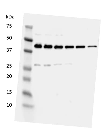
![Western Blot: HMGB1/HMG-1 Antibody [NB100-2322] - Total protein from SHSY-5Y, MCF7, Neuro2A and HeLa was separated on a 12% gel by SDS-PAGE, transferred to PVDF membrane and blocked in 5% non-fat milk in TBST. The membrane was probed with 2.0 ug/mL anti-HMGB1 in 1% non-fat milk in TBST and detected with an anti-rabbit HRP secondary antibody using chemiluminescence.](http://images.novusbio.com/fullsize/HMGB1-HMG-1-Antibody-Western-Blot-NB100-2322-img0017.jpg "Western Blot: HMGB1/HMG-1 Antibody [NB100-2322] - Total protein from SHSY-5Y, MCF7, Neuro2A and HeLa was separated on a 12% gel by SDS-PAGE, transferred to PVDF membrane and blocked in 5% non-fat milk in TBST. The membrane was probed with 2.0 ug/mL anti-HMGB1 in 1% non-fat milk in TBST and detected with an anti-rabbit HRP secondary antibody using chemiluminescence.")
![Immunohistochemistry-Paraffin: HMGB1/HMG-1 Antibody [NB100-2322] - Staining of HMGB1 in mouse liver using NB100-2322.](http://images.novusbio.com/fullsize/HMGB1-HMG-1-Antibody-Immunohistochemistry-Paraffin-NB100-2322-img0011.jpg "Immunohistochemistry-Paraffin: HMGB1/HMG-1 Antibody [NB100-2322] - Staining of HMGB1 in mouse liver using NB100-2322.")
![Simple Western: HMGB1/HMG-1 Antibody [NB100-2322] - Image shows a specific band for HMGB1 in 0.05 mg/mL of Jurkat lysate. This experiment was performed under reducing conditions using the 12-230 kDa separation system.](http://images.novusbio.com/fullsize/HMGB1-HMG-1-Antibody-Simple-Western-NB100-2322-img0014.jpg "Simple Western: HMGB1/HMG-1 Antibody [NB100-2322] - Image shows a specific band for HMGB1 in 0.05 mg/mL of Jurkat lysate. This experiment was performed under reducing conditions using the 12-230 kDa separation system.")
![Immunocytochemistry/Immunofluorescence: HMGB1/HMG-1 Antibody [NB100-2322] - MCF7 cells were fixed in 4% paraformaldehyde for 10 minutes and permeabilized in 0.5% Triton X-100 in PBS for 5 minutes. The cells were incubated with anti-HMGB1/HMG-1 Antibody NB100-2322 at 1 ug/ml for overnight at 4C and detected with an anti-rabbit Dylight 488 (Green) at a 1:1000 dilution for 60 minutes. Nuclei were counterstained with DAPI (Blue). Cells were imaged using a 100X objective and digitally deconvolved.](http://images.novusbio.com/fullsize/HMGB1-HMG-1-Antibody-Immunocytochemistry-Immunofluorescence-NB100-2322-img0027.jpg "Immunocytochemistry/Immunofluorescence: HMGB1/HMG-1 Antibody [NB100-2322] - MCF7 cells were fixed in 4% paraformaldehyde for 10 minutes and permeabilized in 0.5% Triton X-100 in PBS for 5 minutes. The cells were incubated with anti-HMGB1/HMG-1 Antibody NB100-2322 at 1 ug/ml for overnight at 4C and detected with an anti-rabbit Dylight 488 (Green) at a 1:1000 dilution for 60 minutes. Nuclei were counterstained with DAPI (Blue). Cells were imaged using a 100X objective and digitally deconvolved.")
![Immunocytochemistry/Immunofluorescence: HMGB1/HMG-1 Antibody [NB100-2322] - Neuro2a cells were fixed for 10 minutes using 10% formalin and then permeabilized for 5 minutes using 1X TBS + 0.5% Triton-X100. The cells were incubated with anti-HMGB1 at 5 ug/ml overnight at 4C and detected with an anti-rabbit Dylight 488 (Green) at a 1:500 dilution. Alpha tubulin (DM1A) NB100-690 was used as a co-stain at a 1:1000 dilution and detected with an anti-mouse Dylight 550 (Red) at a 1:500 dilution. Nuclei were counterstained with DAPI (Blue). Cells were imaged using a 40X objective.](http://images.novusbio.com/fullsize/HMGB1-HMG-1-Antibody-Immunocytochemistry-Immunofluorescence-NB100-2322-img0018.jpg "Immunocytochemistry/Immunofluorescence: HMGB1/HMG-1 Antibody [NB100-2322] - Neuro2a cells were fixed for 10 minutes using 10% formalin and then permeabilized for 5 minutes using 1X TBS + 0.5% Triton-X100. The cells were incubated with anti-HMGB1 at 5 ug/ml overnight at 4C and detected with an anti-rabbit Dylight 488 (Green) at a 1:500 dilution. Alpha tubulin (DM1A) NB100-690 was used as a co-stain at a 1:1000 dilution and detected with an anti-mouse Dylight 550 (Red) at a 1:500 dilution. Nuclei were counterstained with DAPI (Blue). Cells were imaged using a 40X objective.")
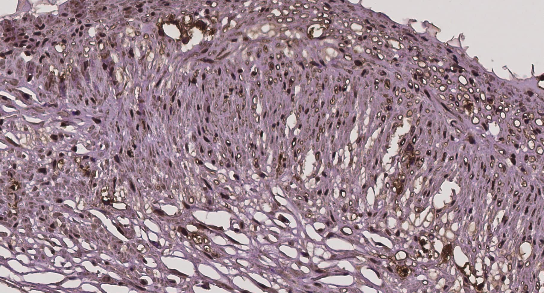
![Immunohistochemistry-Paraffin: HMGB1/HMG-1 Antibody [NB100-2322] - Rabbit blood vessel. Image from verified customer review.](http://images.novusbio.com/fullsize/HMGB1-HMG-1-Antibody-Immunohistochemistry-Paraffin-NB100-2322-img0022.jpg "Immunohistochemistry-Paraffin: HMGB1/HMG-1 Antibody [NB100-2322] - Rabbit blood vessel. Image from verified customer review.")
![Western Blot: HMGB1/HMG-1 Antibody [NB100-2322] - The onset of innate immune response after I/R injury was compared in wt and PT-SSAT-Cko animals. Time course of the expression of HMGB1, TLR2 and 4 were compared in the kidneys of sham-operated and injured wt and PT-SSAT-Cko mice. The data are representative of three independent experiments. Image collected and cropped by CiteAb from the following publication (//doi.org/10.1371/journal.pone.0110161) licensed under a CC-BY license.](http://images.novusbio.com/fullsize/HMGB1-HMG-1-Antibody-Western-Blot-NB100-2322-img0026.jpg "Western Blot: HMGB1/HMG-1 Antibody [NB100-2322] - The onset of innate immune response after I/R injury was compared in wt and PT-SSAT-Cko animals. Time course of the expression of HMGB1, TLR2 and 4 were compared in the kidneys of sham-operated and injured wt and PT-SSAT-Cko mice. The data are representative of three independent experiments. Image collected and cropped by CiteAb from the following publication (//doi.org/10.1371/journal.pone.0110161) licensed under a CC-BY license.")
![Immunocytochemistry/Immunofluorescence: HMGB1/HMG-1 Antibody [NB100-2322] - NIH3T3 cells were fixed in 4% paraformaldehyde for 10 minutes and permeabilized in 0.5% Triton X-100 in PBS for 5 minutes. The cells were incubated with anti-HMGB1/HMG-1 Antibody NB100-2322 at 1 ug/ml for overnight at 4C and detected with an anti-rabbit Dylight 488 (Green) at a 1:1000 dilution for 60 minutes. Nuclei were counterstained with DAPI (Blue). Cells were imaged using a 100X objective and digitally deconvolved.](http://images.novusbio.com/fullsize/HMGB1-HMG-1-Antibody-Immunocytochemistry-Immunofluorescence-NB100-2322-img0028.jpg "Immunocytochemistry/Immunofluorescence: HMGB1/HMG-1 Antibody [NB100-2322] - NIH3T3 cells were fixed in 4% paraformaldehyde for 10 minutes and permeabilized in 0.5% Triton X-100 in PBS for 5 minutes. The cells were incubated with anti-HMGB1/HMG-1 Antibody NB100-2322 at 1 ug/ml for overnight at 4C and detected with an anti-rabbit Dylight 488 (Green) at a 1:1000 dilution for 60 minutes. Nuclei were counterstained with DAPI (Blue). Cells were imaged using a 100X objective and digitally deconvolved.")
![Flow Cytometry: HMGB1/HMG-1 Antibody [NB100-2322] - An intracellular stain was performed on HeLa cells with HMGB1/HMG-1 Antibody NB100-2322AF488 (blue) and a matched isotype control (orange). Cells were fixed with 4% PFA and then permeabilized with 0.1% saponin. Cells were incubated in an antibody dilution of 5 ug/mL for 30 minutes at room temperature. Both antibodies were conjugated to Alexa Fluor 488.](http://images.novusbio.com/fullsize/HMGB1-HMG-1-Antibody-Flow-Cytometry-NB100-2322-img0029.jpg "Flow Cytometry: HMGB1/HMG-1 Antibody [NB100-2322] - An intracellular stain was performed on HeLa cells with HMGB1/HMG-1 Antibody NB100-2322AF488 (blue) and a matched isotype control (orange). Cells were fixed with 4% PFA and then permeabilized with 0.1% saponin. Cells were incubated in an antibody dilution of 5 ug/mL for 30 minutes at room temperature. Both antibodies were conjugated to Alexa Fluor 488.")
![Western Blot: HMGB1/HMG-1 Antibody [NB100-2322] - Hepatocyte protein lysate at 1:1000 4C overnight. Image from verfified customer review.](http://images.novusbio.com/fullsize/HMGB1-HMG-1-Antibody-Western-Blot-NB100-2322-img0013.jpg "Western Blot: HMGB1/HMG-1 Antibody [NB100-2322] - Hepatocyte protein lysate at 1:1000 4C overnight. Image from verfified customer review.")
![Immunocytochemistry/Immunofluorescence: HMGB1/HMG-1 Antibody [NB100-2322] - HeLa cells were fixed for 10 minutes using 10% formalin and then permeabilized for 5 minutes using 1X TBS + 0.5% Triton-X100. The cells were incubated with anti-HMGB1 [NB100-2322] at a 1:200 dilution overnight at 4C and detected with an anti-rabbit Dylight 488 (Green) at a 1:500 dilution. Alpha tubulin (DM1A) [NB100-690] was used as a co-stain at a 1:1000 dilution and detected with an anti-mouse Dylight 550 (Red) at a 1:500 dilution. Nuclei were counterstained with DAPI (Blue). Cells were imaged using a 40X objective.](http://images.novusbio.com/fullsize/HMGB1-HMG-1-Antibody-Immunocytochemistry-Immunofluorescence-NB100-2322-img0015.jpg "Immunocytochemistry/Immunofluorescence: HMGB1/HMG-1 Antibody [NB100-2322] - HeLa cells were fixed for 10 minutes using 10% formalin and then permeabilized for 5 minutes using 1X TBS + 0.5% Triton-X100. The cells were incubated with anti-HMGB1 [NB100-2322] at a 1:200 dilution overnight at 4C and detected with an anti-rabbit Dylight 488 (Green) at a 1:500 dilution. Alpha tubulin (DM1A) [NB100-690] was used as a co-stain at a 1:1000 dilution and detected with an anti-mouse Dylight 550 (Red) at a 1:500 dilution. Nuclei were counterstained with DAPI (Blue). Cells were imaged using a 40X objective.")
![Flow (Intracellular): HMGB1/HMG-1 Antibody [NB100-2322] - An intracellular stain was performed on HeLa with NB100-2322 and a matched isotype control. Cells were fixed with 4% PFA and then permeabilized with 0.1% saponin. Cells were incubated in an antibody dilution of 1 ug/mL for 30 minutes at room temperature, followed by Rabbit IgG (H+L) Cross-Adsorbed Secondary Antibody.](http://images.novusbio.com/fullsize/HMGB1-HMG-1-Antibody-Flow-Intracellular-NB100-2322-img0021.jpg "Flow (Intracellular): HMGB1/HMG-1 Antibody [NB100-2322] - An intracellular stain was performed on HeLa with NB100-2322 and a matched isotype control. Cells were fixed with 4% PFA and then permeabilized with 0.1% saponin. Cells were incubated in an antibody dilution of 1 ug/mL for 30 minutes at room temperature, followed by Rabbit IgG (H+L) Cross-Adsorbed Secondary Antibody.")
![Flow Cytometry: HMGB1/HMG-1 Antibody [NB100-2322] - An intracellular stain was performed on RH-30 cells with NB100-2322F (blue) and a matched isotype control (orange). Cells were fixed with 4% PFA and then permeabilized with 0.1% saponin. Cells were incubated in an antibody dilution of 10 ug/mL for 30 minutes at room temperature. Both antibodies were conjugated to FITC.](http://images.novusbio.com/fullsize/HMGB1-HMG-1-Antibody-Flow-Cytometry-NB100-2322-img0024.jpg "Flow Cytometry: HMGB1/HMG-1 Antibody [NB100-2322] - An intracellular stain was performed on RH-30 cells with NB100-2322F (blue) and a matched isotype control (orange). Cells were fixed with 4% PFA and then permeabilized with 0.1% saponin. Cells were incubated in an antibody dilution of 10 ug/mL for 30 minutes at room temperature. Both antibodies were conjugated to FITC.")
![ELISA: HMGB1/HMG-1 Antibody [NB100-2322] - A dose-dependent titration of the HRP conjugated anti-HMGB1 antibody on recombinant human HMGB1 protein. Image from verified customer review. Image using the HRP format of this antibody.](http://images.novusbio.com/fullsize/HMGB1-HMG-1-Antibody-ELISA-NB100-2322-img0023.jpg "ELISA: HMGB1/HMG-1 Antibody [NB100-2322] - A dose-dependent titration of the HRP conjugated anti-HMGB1 antibody on recombinant human HMGB1 protein. Image from verified customer review. Image using the HRP format of this antibody.")
 Expression of biglycan in AD-TLR2KO mice increased significantly compared with that in WT, AD, and TLR2KO mice (p0.05).")
![Western Blot: HMGB1/HMG-1 Antibody [NB100-2322] - Expression of endogenous ligands for TLR2. (A) Expression of biglycan in AD-TLR2KO mice increased significantly compared with that in WT, AD, & TLR2KO mice (p0.05). Image collected & cropped by CiteAb from the following publication (//pubmed.ncbi.nlm.nih.gov/31509519), licensed under a CC-BY license. Not internally tested by Novus Biologicals.](http://images.novusbio.com/fullsize/nb100-2322_rabbit-polyclonal-hmgb1-hmg-1-antibody-310202415175213.jpg "Western Blot: HMGB1/HMG-1 Antibody [NB100-2322] - Expression of endogenous ligands for TLR2. (A) Expression of biglycan in AD-TLR2KO mice increased significantly compared with that in WT, AD, & TLR2KO mice (p0.05). Image collected & cropped by CiteAb from the following publication (//pubmed.ncbi.nlm.nih.gov/31509519), licensed under a CC-BY license. Not internally tested by Novus Biologicals.")
![SDS-PAGE TNF-alpha [Unconjugated]](https://images.novusbio.com/images2/TNF-alpha_210-TA_256.jpg)
![Bioactivity TNF-alpha [Unconjugated]](https://images.novusbio.com/images2/TNFalpha_210TA_1658.jpg)
![SEC-MALS TNF-alpha [Unconjugated]](https://images.novusbio.com/images/210-ta_recombinant-human-tnf-alpha-protein-sec-mals-35202312244..jpg)
![N/A IL-6 [HRP]](https://images.novusbio.com/images2/DATA_IL6_M6000_ELISA_936.jpg)
![N/A IL-6 [HRP]](https://images.novusbio.com/images2/IL-6_M6000_ELISA_415.jpg)
![N/A IL-6 [HRP]](https://images.novusbio.com/images/m6000b_mouse-il-6-quantikine-elisa-kit-44202415433118.jpg)
![N/A AGER [HRP]](https://images.novusbio.com/images2/DATA_RAGE_DRG00_ELISA_806.jpg)
![N/A AGER [HRP]](https://images.novusbio.com/images2/DATA_RAGE_DRG00_ELISA_807.jpg)
![N/A AGER [HRP]](https://images.novusbio.com/images2/RAGE_DRG00_ELISA_171.jpg)
![N/A IL-10 [Biotin]](https://images.novusbio.com/images2/DATA_IL10_DY417_ELISA_2014.jpg)
![Western Blot ERK2 Antibody [Unconjugated]](https://images.novusbio.com/images2/ERK2_AF1230_Western_Blot_5097.jpg)
![Immunohistochemistry ERK2 Antibody [Unconjugated]](https://images.novusbio.com/images2/ERK2_AF1230_Immunohistochemistry_20696.jpg)
![Knockout Validated ERK2 Antibody [Unconjugated]](https://images.novusbio.com/images2/ERK2_AF1230_Knockout_Validated_22864.jpg)


![N/A CCL2/JE/MCP-1 [HRP]](https://images.novusbio.com/images2/CCL2_DCP00_ELISA_67.jpg)
![N/A CCL2/JE/MCP-1 [HRP]](https://images.novusbio.com/images2/DATA_CCL2_DCP00_ELISA_633.jpg)
![N/A CCL2/JE/MCP-1 [HRP]](https://images.novusbio.com/images2/DATA_CCL2_DCP00_ELISA_634.jpg)
![Western Blot: Goat anti-Rabbit IgG (H+L) Secondary Antibody [HRP] [NB7160] - Western blot showing vemurafenib treatment in BRAFV600E CRC cells inhibits fission mediator DRP1 with no significant effect on fusion proteins (Mfn1 & 2) using MFN-1 antibody (NBP1-51841) and corresponding secondary antibody, goat anti-rabbit IgG-HRP (NB7160). Image collected and cropped by CiteAb from the following publication (https://pubmed.ncbi.nlm.nih.gov/33738242).](https://images.novusbio.com/images/Goat-anti-Rabbit-IgG-H+L-Secondary-Antibody-HRP-Western-Blot-NB7160-img0001.jpg "Western Blot: Goat anti-Rabbit IgG (H+L) Secondary Antibody [HRP] [NB7160] - Western blot showing vemurafenib treatment in BRAFV600E CRC cells inhibits fission mediator DRP1 with no significant effect on fusion proteins (Mfn1 & 2) using MFN-1 antibody (NBP1-51841) and corresponding secondary antibody, goat anti-rabbit IgG-HRP (NB7160). Image collected and cropped by CiteAb from the following publication (https://pubmed.ncbi.nlm.nih.gov/33738242).")
 using a 1:1000 dilution of HRP-conjugated Anti-Rabbit IgG Secondary Antibody (Catalog # HAF008). This experiment was conducted under reducing conditions and using Immunoblot Buffer Group 1.")
![Flow Cytometry: Rabbit IgG Isotype Control [NBP2-24891] - Intracellular FACS analysis of mouse TLR6 polyclonal antibody (red), rabbit isotype control (green), RAW cells alone (shaded). Two micrograms of antibodies were used. Goat anti-rabbit FITC (Novus, 20302) was used as secondary.](https://images.novusbio.com/images/Rabbit--Mouse-IgG-Isotype-Control-Flow-Cytometry-NBP2-24891-img0001.jpg "Flow Cytometry: Rabbit IgG Isotype Control [NBP2-24891] - Intracellular FACS analysis of mouse TLR6 polyclonal antibody (red), rabbit isotype control (green), RAW cells alone (shaded). Two micrograms of antibodies were used. Goat anti-rabbit FITC (Novus, 20302) was used as secondary.")


